


Vol 2, No 1 (2026): Current Issue
Original Article
Review Articles
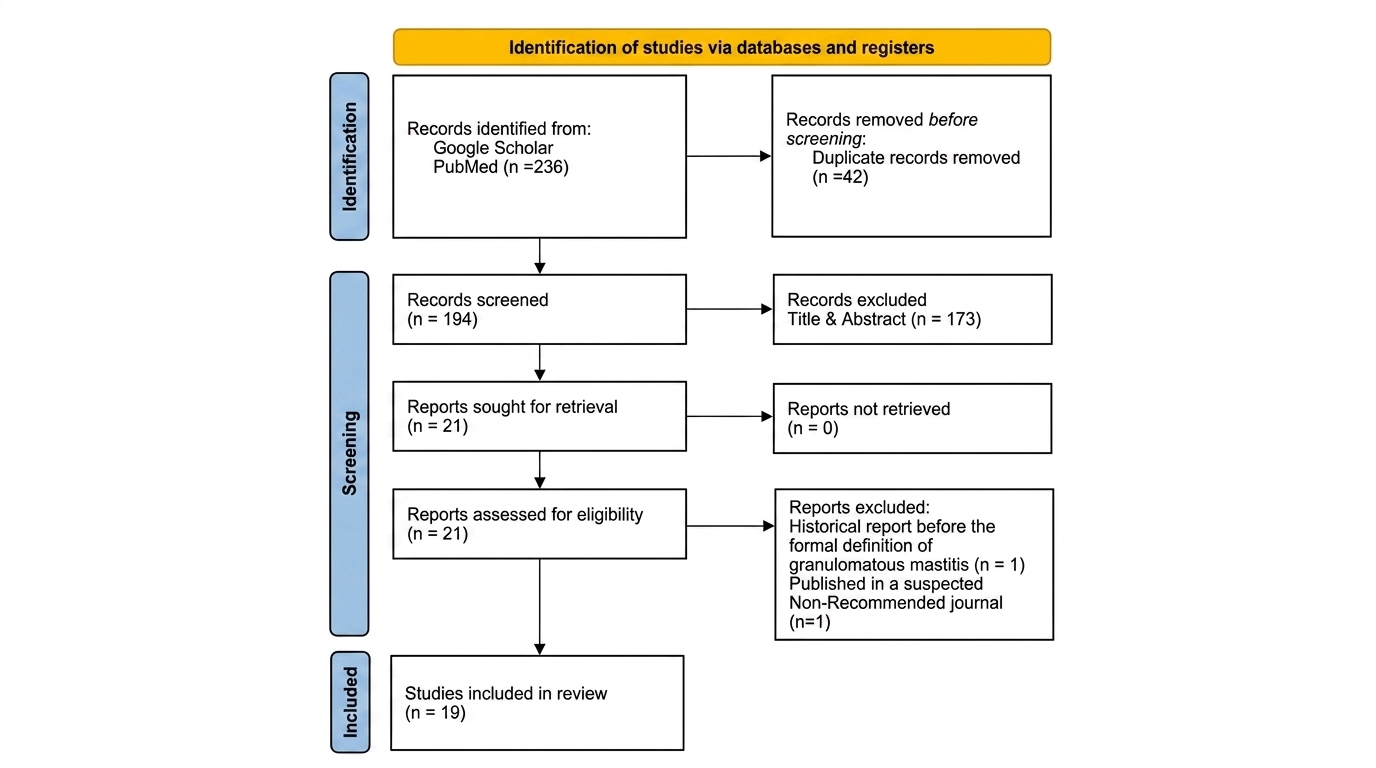
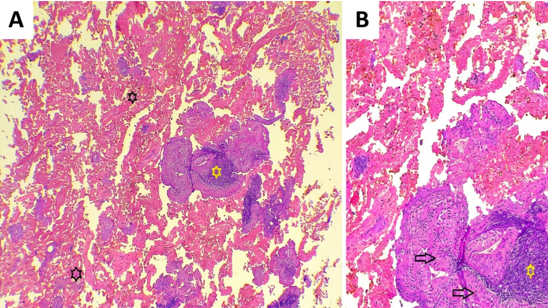
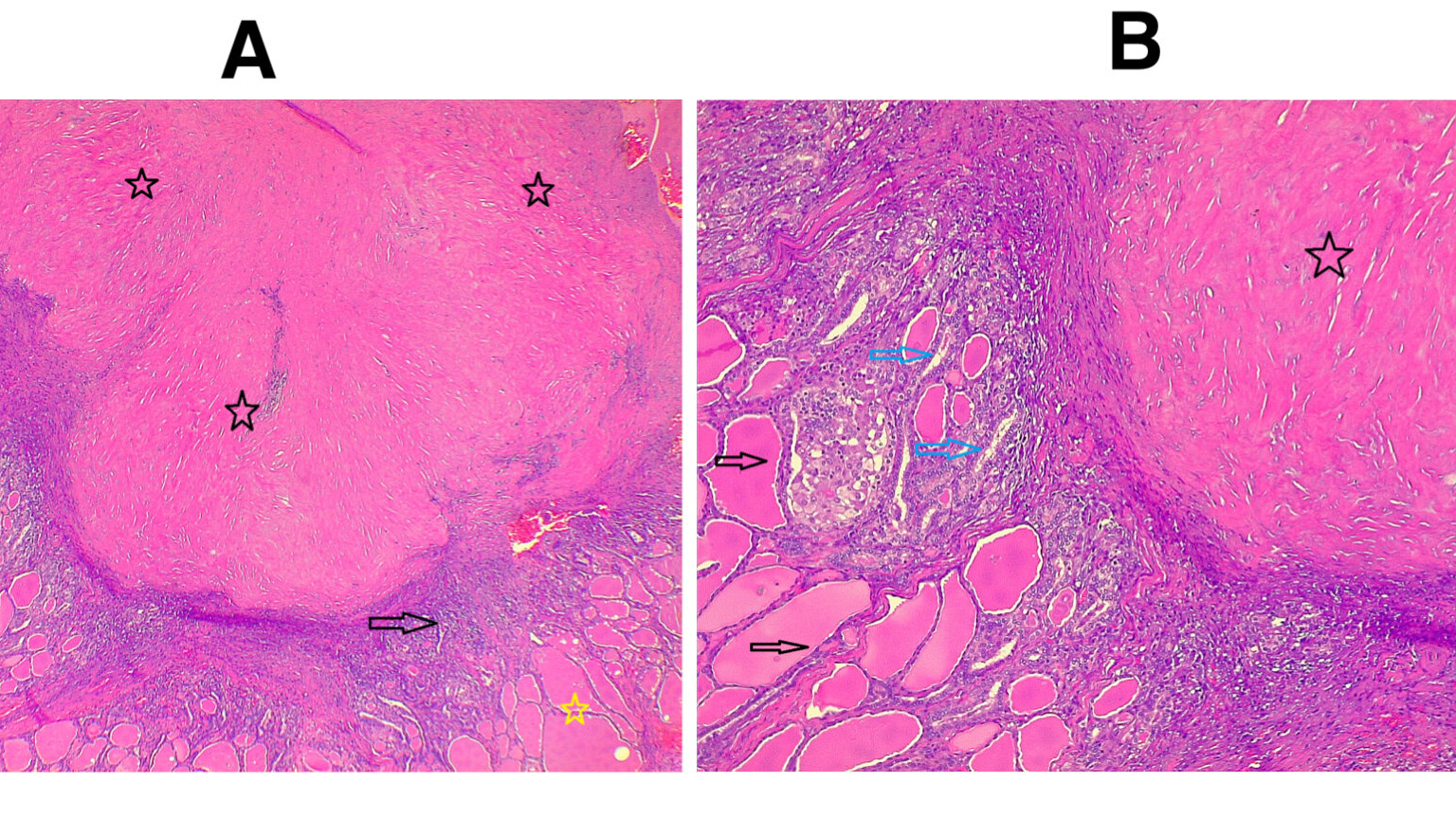
Case Reports
Judi Clinical Journal is a leading open-access journal that covers a wide range of clinical sciences. The journal is dedicated to publishing significant advancements in clinical research that are of importance to specialists across various medical disciplines.
We are committed to providing a streamlined and efficient service for both authors and readers. Our independent editorial team ensures timely and fair publication decisions. Accepted manuscripts are swiftly disseminated to a broad audience through our continuous online publication model, enabling rapid and widespread access to the latest clinical research findings.
We accept submissions including original research, systematic reviews, meta-analyses, review articles, case reports, case series, editorials, letters to the editor, commentaries, perspectives, short communications, and correspondence. The journal primarily focuses on the following areas:
- Evidence-based medicine.
- Public health and healthcare policies.
- Current diseases’ diagnosis and management.
- Clinical pharmacology
- Pathology
- Radiology
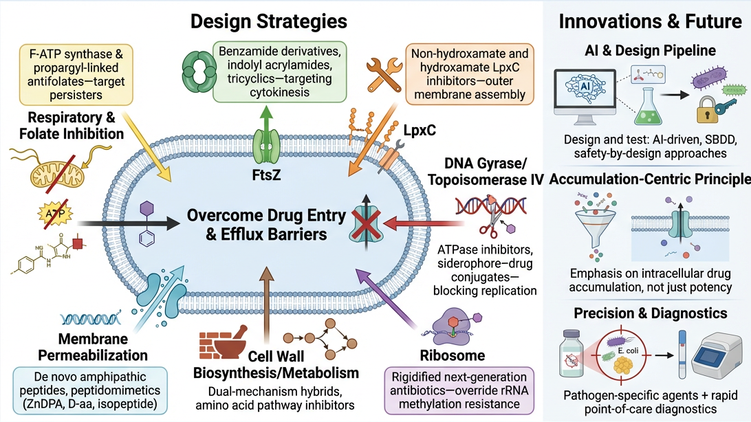
- Clinical and applied studies like surgery and innovated techniques, Anesthesiology, Cardiology, Dermatology, Emergency Medicine, Endocrinology, Family Medicine, Gastroenterology, General Surgery, Geriatrics, Gynecology, Hematology, Infectious Diseases, Internal Medicine, Medical Genetics, Nephrology, Neurology, Neurosurgery, Obstetrics, Oncology, Ophthalmology, Orthopedic Surgery, Otolaryngology (ENT), Palliative Care, Pediatrics, Physical Medicine and Rehabilitation, Plastic Surgery, Psychiatry, Pulmonology, Rheumatology, Sports Medicine, Thoracic Surgery, Urology, Vascular Surgery.
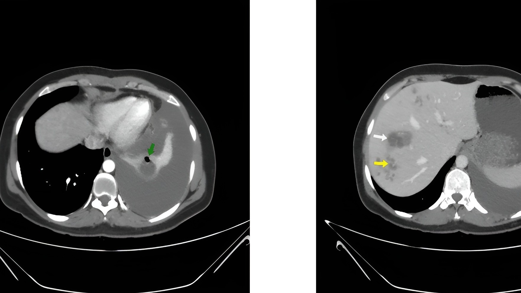
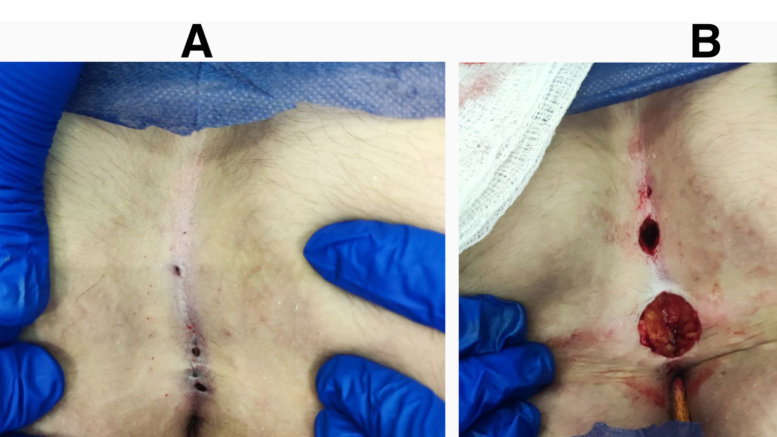
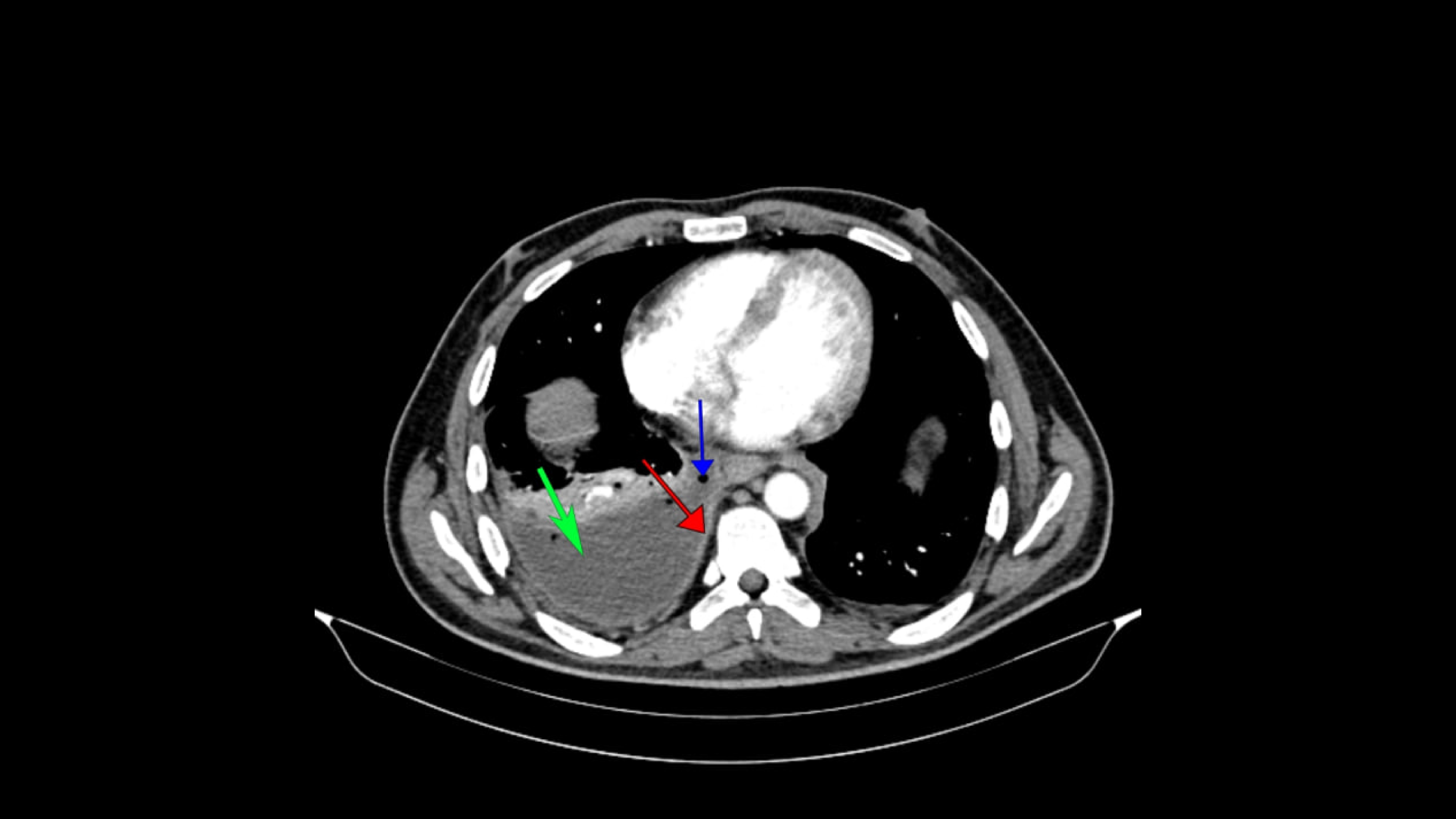
Latest Articles