Shaho F. Ahmed, Rebaz M. Ali, Rawa M. Ali, Ari M. Abdullah, Abdulwahid M. Salih, Hiwa O. Baba, Rebaz O. Mohammed, Aso N. Qadir, Shko H. Hassan, Abdullah A. Qadir, Harun A. Ahmed, Hawkar A. Nasralla, Berun A. Abdalla, Fahmi H. Kakamad (Author)
Preoperative Thyroglobulin and Thyroid Pathologies: A Single Center Experience
Shaho F. Ahmed, Rebaz M. Ali, Rawa M. Ali, Ari M. Abdullah, Abdulwahid M. Salih, Hiwa O....
Introduction: Thyroid nodules are frequently found in the general population, though malignancy is confirmed in only a minority of cases. Distinguishing between benign and malignant nodules before surgery is vital for appropriate clinical management. The utility of preoperative serum thyroglobulin (Tg) levels as a diagnostic marker in thyroid carcinomas remains controversial. This study aimed to provide a descriptive overview of patients with markedly elevated preoperative Tg levels (>500 ng/mL) and their corresponding histopathological outcomes.
Methods: A retrospective, single-center study was conducted at Smart Health Tower, which included patients who underwent surgical interventions for thyroid disorders between 2019 and 2025, with preoperative serum Tg levels exceeding 500 ng/mL. Patients were excluded if they had incomplete medical records. Patient demographics, clinical features, preoperative findings, surgical details, and final histopathology were retrieved from electronic medical records.
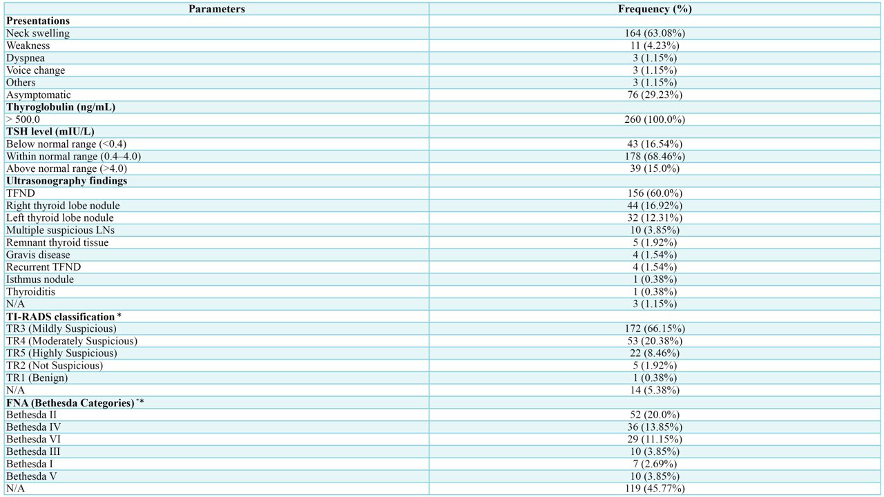
Results: A total of 260 patients were included, predominantly female (73.08%), with a median age of 49 years (QR: 38–61). Neck swelling was the most common symptom (63.08%). Ultrasonography showed follicular nodular disease in 60.0%. Fine-needle aspiration cytology revealed Bethesda II in 20.0%, IV in 13.85%, and VI in 11.15% of cases. Total thyroidectomy was performed in 74.62% of cases. Histopathology showed benign lesions in 70.77% and malignant lesions in 29.23% of the cases.
Conclusion: Preoperative Tg levels may be elevated in both benign and malignant thyroid disorders; however, Tg may not possess adequate diagnostic precision to be used as a sole indicator of malignancy.
Shaban Latif, Hemn H. Kaka Ali, Deari A. Ismaeil, Karzan M. Salih, Ayman M. Mustafa, Shko H. Hassan, Kayhan A. Najar, Harun A. Ahmed, Abdullah A. Qadir, Bushra O. Hussein (Author)
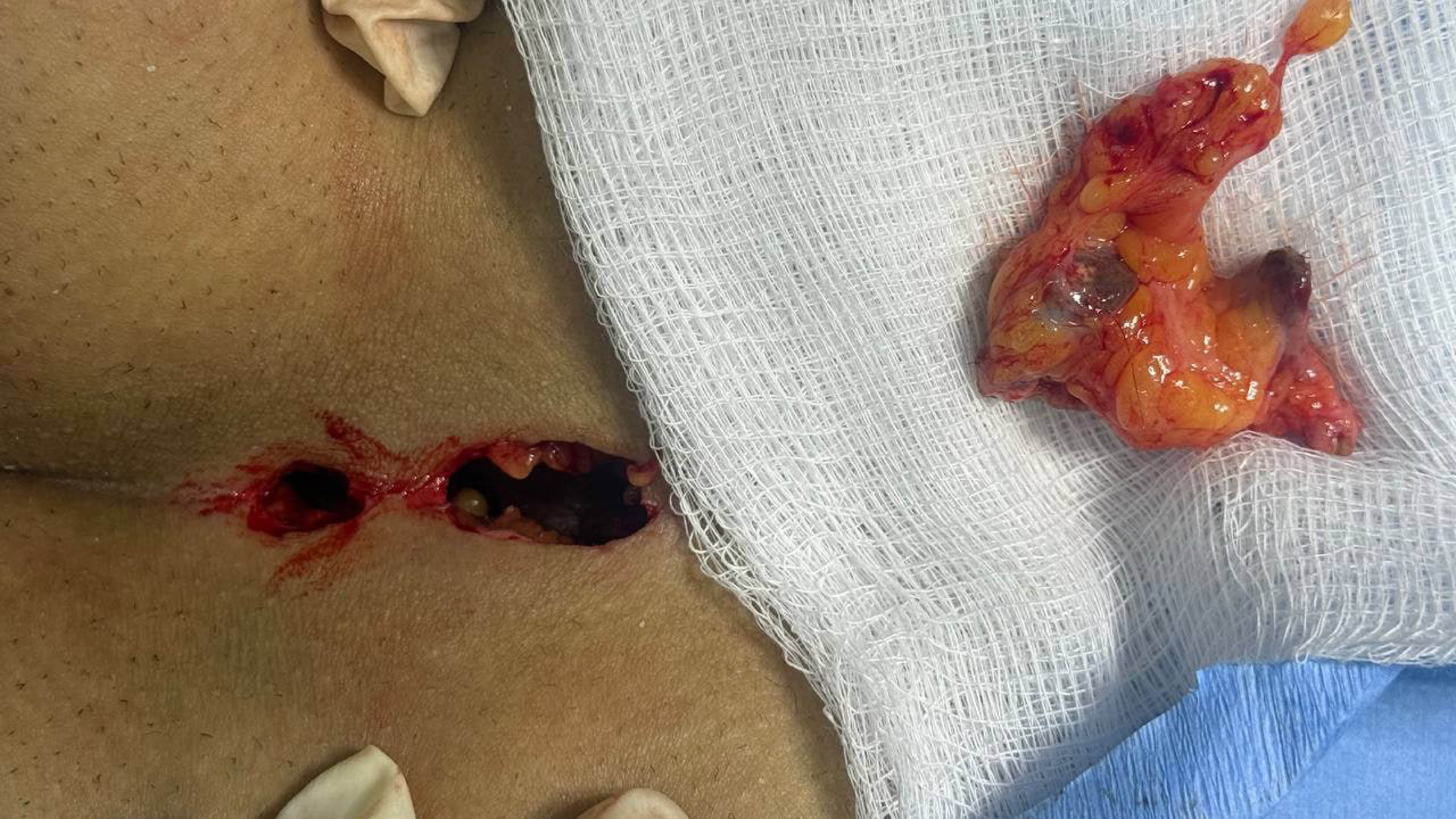
Atypical Presentations of Pilonidal Sinus Disease: A Case Series with Literature Review
Shaban Latif, Hemn H. Kaka Ali, Deari A. Ismaeil, Karzan M. Salih, Ayman M. Mustafa, Shko H....
Introduction: Pilonidal sinus (PNS) typically arises in the sacrococcygeal region but can occasionally present in atypical locations, including the axilla, intermammary region, umbilicus, interdigital web, neck, endoanal, auricular, periauricular, scalp, and submental areas. This study aims to examine clinical features, surgical management, and outcomes of PNS in these uncommon sites.
Methods: This retrospective case series was conducted at a single center over eight years and examined patients with extra-sacrococcygeal PNS confirmed by histopathology. Clinical, surgical, and follow-up data were retrieved from patient records. Data were analyzed using SPSS 25.0 and presented as descriptive statistics (frequencies, percentages, means, and ranges).
Results: This study included 4 patients, with an equal sex distribution (2 females, 50%; 2 males, 50%) and a mean age of 24 years (range: 16–31). Presenting symptoms included discharge, itching, pain, redness, and mass formation. None had significant past medical history, and one patient (25%) had prior surgery at the same site. Affected sites were equally distributed: breast (25%), axilla (25%), penile (25%), and scalp (25%). All patients underwent surgical intervention, and histopathology revealed tracts lined with epidermis, surrounded by mixed inflammatory cells, foreign body giant cells, and hair shafts in granulation tissue. During the follow-up period (mean follow-up: 13.5 months), no recurrence was observed.
Conclusion: Atypical presentations of PNS, although rare, can occur in diverse anatomical regions. Surgical excision with primary closure might be an effective treatment approach, potentially leading to favorable clinical outcomes and a low recurrence rate.
Abdulwahid M. Salih, Lana RA. Pshtiwan, Fattah H. Fattah, Ari M. Abdullah, Rebaz M. Ali, Rebaz O. Mohammed, Hiwa O. Baba, Ahmed H. Ahmed, Sakar O. Arif, Nasren S. Sabr, Shko H. Hassan, Harzal H. Fatih, Masty K. Ahmed, Meer M. Abdulkarim, Hiwa O. Abdullah, Fahmi H. Kakamad (Author)
Abdulwahid M. Salih, Lana RA. Pshtiwan, Fattah H. Fattah, Ari M. Abdullah, Rebaz M. Ali, Rebaz...
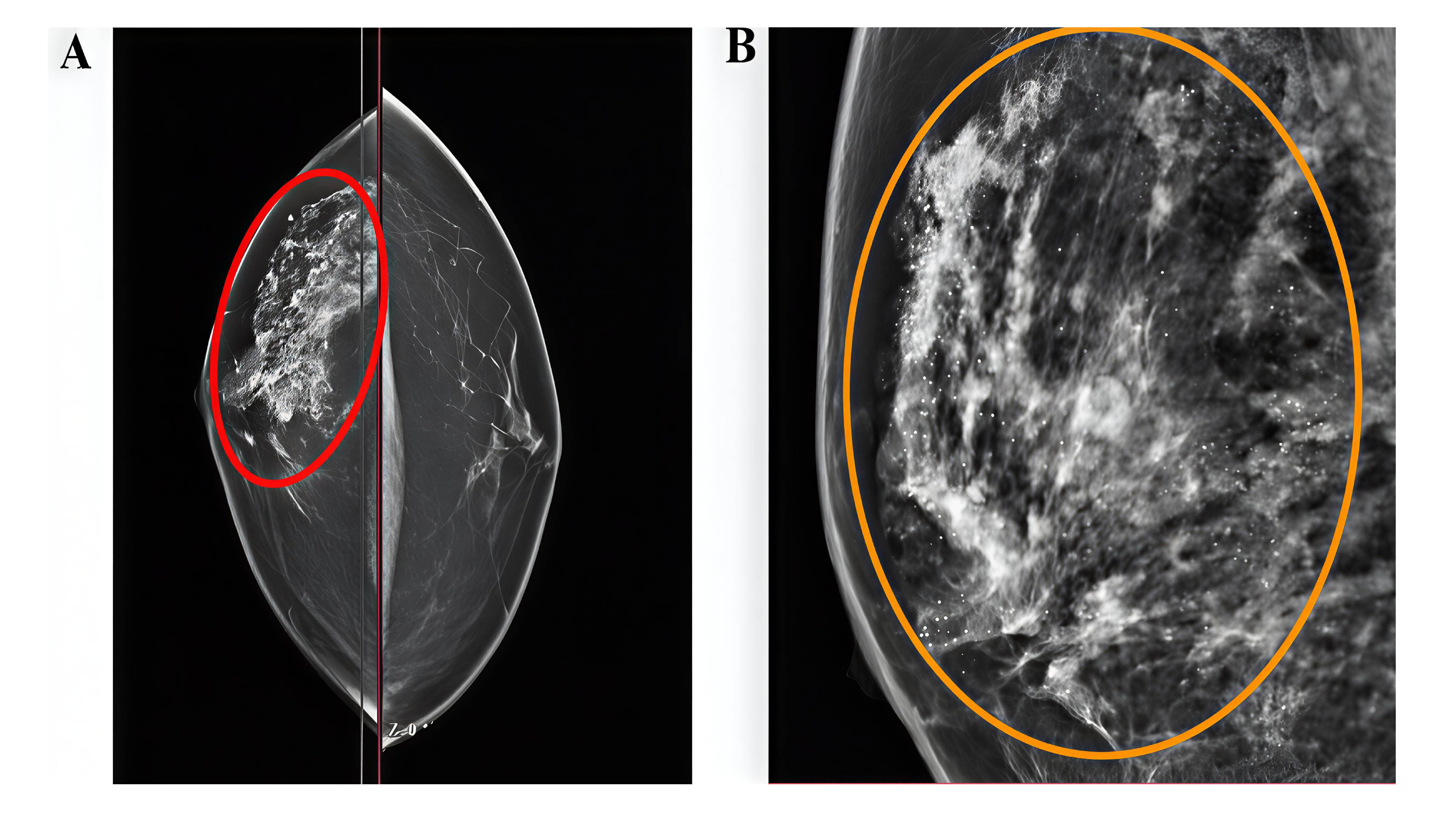
Introduction: Male breast cancer is a rare malignancy, and despite increasing its prevalence, little is known about its prognosis, and management continues to rely largely on guidelines developed for female breast cancer. This study presents a single-center experience focusing on the diagnosis, therapeutic strategies, and prognostic outcomes associated with male breast cancer.
Methods: The study was carried out between May 2020 and January 2025. All the male patients diagnosed with breast cancer were identified from the center's database. The study included those with complete medical records, encompassing demographics, clinical presentation, imaging findings, outcome, and follow-up data.
Results: Fifteen male breast cancer cases were analyzed (mean age 59.47 ± 12.97 years). The left breast was affected in 60%, and 80% presented with a painless lump. Tumor size ranged 0.6–5.4 cm (mean 2.53 ± 1.50 cm). Modified radical mastectomy was performed in 86.66%. Invasive ductal carcinoma predominated (93.33%), with lymph node involvement in 40% and bone metastasis in 6.67%. Immunohistochemistry results were available for 5 patients (33.33%); all of them were estrogen receptor and progesterone receptor positive and human epidermal growth factor receptor 2 negative. Adjuvant chemotherapy and radiotherapy were used in 33.33%, while 20% received triple modality therapy. Mean follow-up was 2.8 ± 1.52 years (range 1–5), with one recurrence.
Conclusion: Male breast cancer may present as a breast mass and is often diagnosed as invasive ductal carcinoma. Modified radical mastectomy accompanied by adjuvant therapy might offer favorable outcomes.
Fahmi H. Kakamad, Berun A. Abdalla, Dahat A. Hussein, Fakher Abdullah, Ayman M. Mustafa, Sabah J. Hasan, Mohammed S. Ezzat, Harem S. Amin, Lanya Q. Rashid, Muhammad A. Khdir, Danyar G. HamaSaeed, Hussein H. Rasul, Akam R. Ahmed (Author)
Clinicopathological Profile of Malignant Tumors at a Tertiary Center in Iraq: A Five-Year Analysis
Fahmi H. Kakamad, Berun A. Abdalla, Dahat A. Hussein, Fakher Abdullah, Ayman M. Mustafa,...
Introduction: Cancer incidence and mortality are increasing worldwide, disproportionately affecting low- and middle-income countries due to limited healthcare access. This study aims to analyze the prevalence and clinicopathological characteristics of cancers diagnosed at a tertiary care center, offering crucial data to inform regional cancer control efforts.
Methods: This retrospective study was conducted at a single tertiary center, analyzing histopathological reports from January 2020 to January 2025. Data were collected from digital and physical archives by a trained team, including only confirmed malignant diagnoses. Variables extracted included patient demographics, cancer types, tumor characteristics, and procedure details. Data integrity was ensured through double-entry and expert review. Statistical analysis using SPSS 27.0 summarized categorical variables as frequencies, percentages and continuous variables as means or medians.
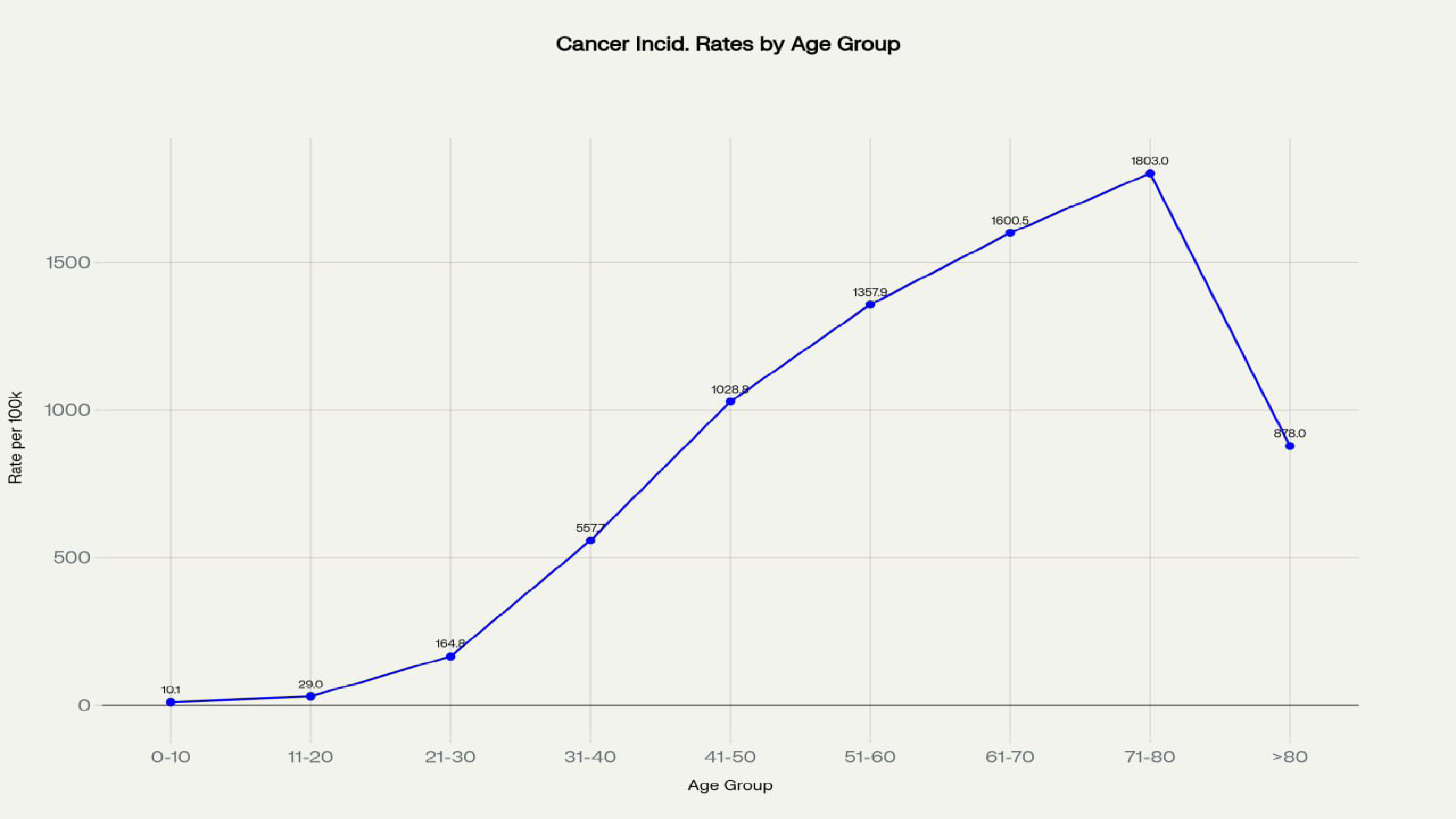
Results: A total of 9,375 cases were analyzed (median age 48 years), with females comprising 75.74% (7,101 cases). The age-standardized incidence rate was 82.20 per 100,000. Head and neck cancers accounted for 2,903 cases (30.97%), primarily thyroid (2,208 cases), followed by thoracic malignant tumors with 2,556 cases (27.26%). Invasive ductal carcinoma was most frequent cancer type (2,392 cases, 25.5%). Larger tumors correlated significantly with positive margins and lymph node involvement (p < 0.05).
Conclusion: The study highlights a unique cancer profile, with younger age at diagnosis and higher rates of head, neck, and thyroid cancers. These findings underscore the need for targeted screening and prevention strategies adapted to regional healthcare systems.
Introduction: A collision tumor consists of two distinct neoplastic components located within the same organ, separated by stromal tissue, without histological intermixing. These rare tumors are usually identified incidentally in surgical specimens. This study systematically reviews complex collision tumors (those with three or more distinct histological types) to explore their features and clinical behavior.
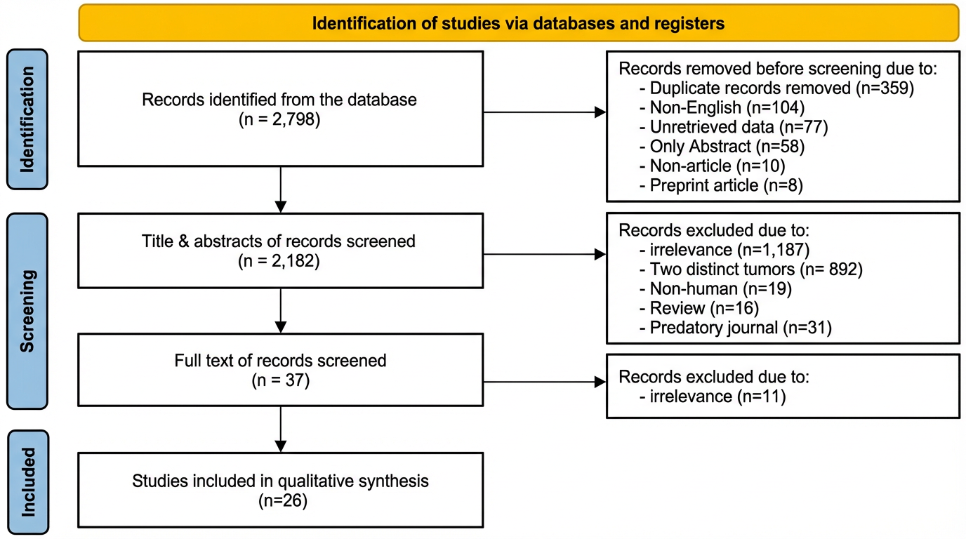
Methods: A comprehensive literature search was conducted using Google Scholar, Consensus AI, and Perplexity AI to identify all articles that describe collision tumors comprising more than two distinct pathologies. Studies lacking full texts, reviews, or those from predatory journals were excluded. Data extracted included publication details, patient demographics, clinical and diagnostic findings, tumor characteristics, treatments, outcomes, and follow-up. Findings were analyzed qualitatively and summarized using frequencies, percentages, and means with standard deviations.
Results: A total of 2,798 articles were identified, and 26 studies (28 cases) met the inclusion criteria. Female patients accounted for 17 cases (60.72%), with a mean age of 63.46 ± 14.00 years. Surgery was performed in 26 cases (92.86%). Triple collision tumors were reported in 26 cases (92.86%), and quadruple collision tumors in 2 cases (7.14%). The thyroid gland was affected in 7 cases (25.00%), and papillary thyroid carcinoma was identified in 9 cases (32.14%). At the last follow-up, 22 patients (78.57%) were alive.
Conclusion: Complex collision tumors represent rare and histologically diverse entities with significant diagnostic and therapeutic implications. They are most frequently found in the thyroid and skin. Accurate diagnosis typically requires comprehensive histopathological and immunohistochemical analysis of the entire lesion.
Ronak S. Ahmed, Shvan O. Siddiq, Lawen J. Mustafa, Rawa M. Ali, Razan B. Jalal, Omed M. Hussein, Saywan K. Asaad, Abdullah K. Ghafour, Lawand A. Sharif, Sasan M. Ahmed (Author)
Late-Onset Alkaptonuria in an Elderly Male: A Case Report and Literature Review
Ronak S. Ahmed, Shvan O. Siddiq, Lawen J. Mustafa, Rawa M. Ali, Razan B. Jalal, Omed M....
Introduction: Alkaptonuria (AKU) is a rare autosomal recessive metabolic disorder caused by homogentisate 1,2-dioxygenase deficiency, leading to homogentisic acid accumulation. While AKU typically presents in children and young adults, this report aims to present an unusual late-onset presentation of AKU in an elderly male.
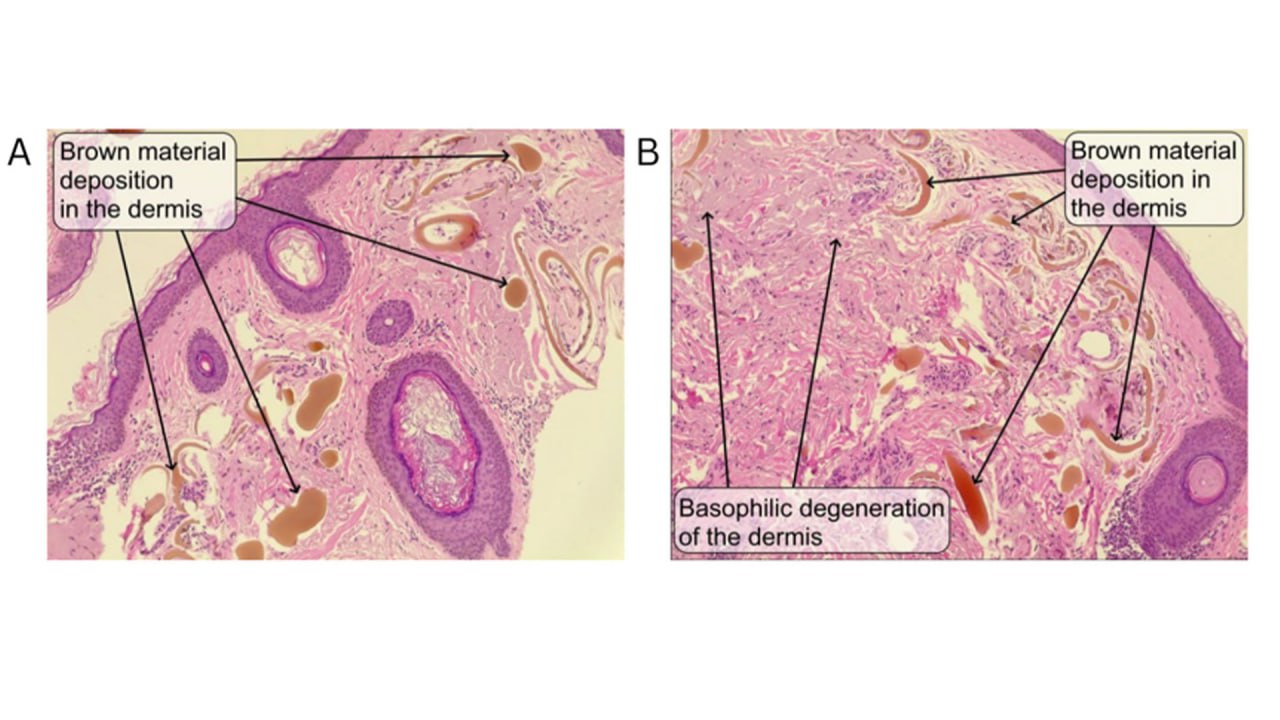
Case presentation: A 65-year-old male presented with 20 years of black skin discoloration and chronic knee and pelvic pain. Despite early signs, including dark-colored urine, the condition was repeatedly misdiagnosed as melasma and primary osteoarthritis. Computed tomography scan revealed vertebral ankylosis and sacroiliac arthritis, and histopathological examination confirmed ochronotic pigment deposition. Due to financial constraints, the patient declined joint replacement surgery and was managed with diclofenac 100 mg twice daily and dietary restrictions.
Literature review: Among six case reports of AKU reviewed in the literature, selected with a focus on misdiagnosis or delayed diagnosis, the majority of cases (3/6, 50%) occurred in the 8th–9th decades of life. Although AKU affects both sexes equally, five of the six reviewed cases (83.3%) were male.
Conclusion: This case highlights the unusual late-onset alkaptonuria and the risk of prolonged misdiagnosis, emphasizing the importance of early recognition and multidisciplinary management.
Yadgar N. Abbas, Fattah H. Fattah, Hemin S. Mohammed, Meer M. Abdulkarim, Jihad I. Hama, Amr M. Mahmood, Hussein M. Hamasalih, Shvan H. Mohammed, Berun A. Abdalla (Author)
Fregoli Syndrome: A Case Report and Literature Review
Yadgar N. Abbas, Fattah H. Fattah, Hemin S. Mohammed, Meer M. Abdulkarim, Jihad I. Hama, Amr...
Introduction: Fregoli syndrome is a rare misidentification disorder that can disrupt behavior, endanger safety, and impair quality of life. Its occurrence in young adults is exceptionally uncommon. This report presents a case of Fregoli delusion in a young adult without the usual underlying causes, such as schizophrenia.
Case presentation: A 25-year-old male presented with a two-month history of persecutory delusions, believing that strangers, friends, and professors were disguising themselves to follow and harass him. His psychiatric history was unremarkable, and there was no family history of mental illness. The patient was diagnosed with Fregoli delusion and prescribed Risperidone 2 mg/day, alongside cognitive-behavioral therapy to challenge his delusions. After three months of treatment, he showed gradual improvement, with reduced intensity of delusions but occasional paranoid thoughts.
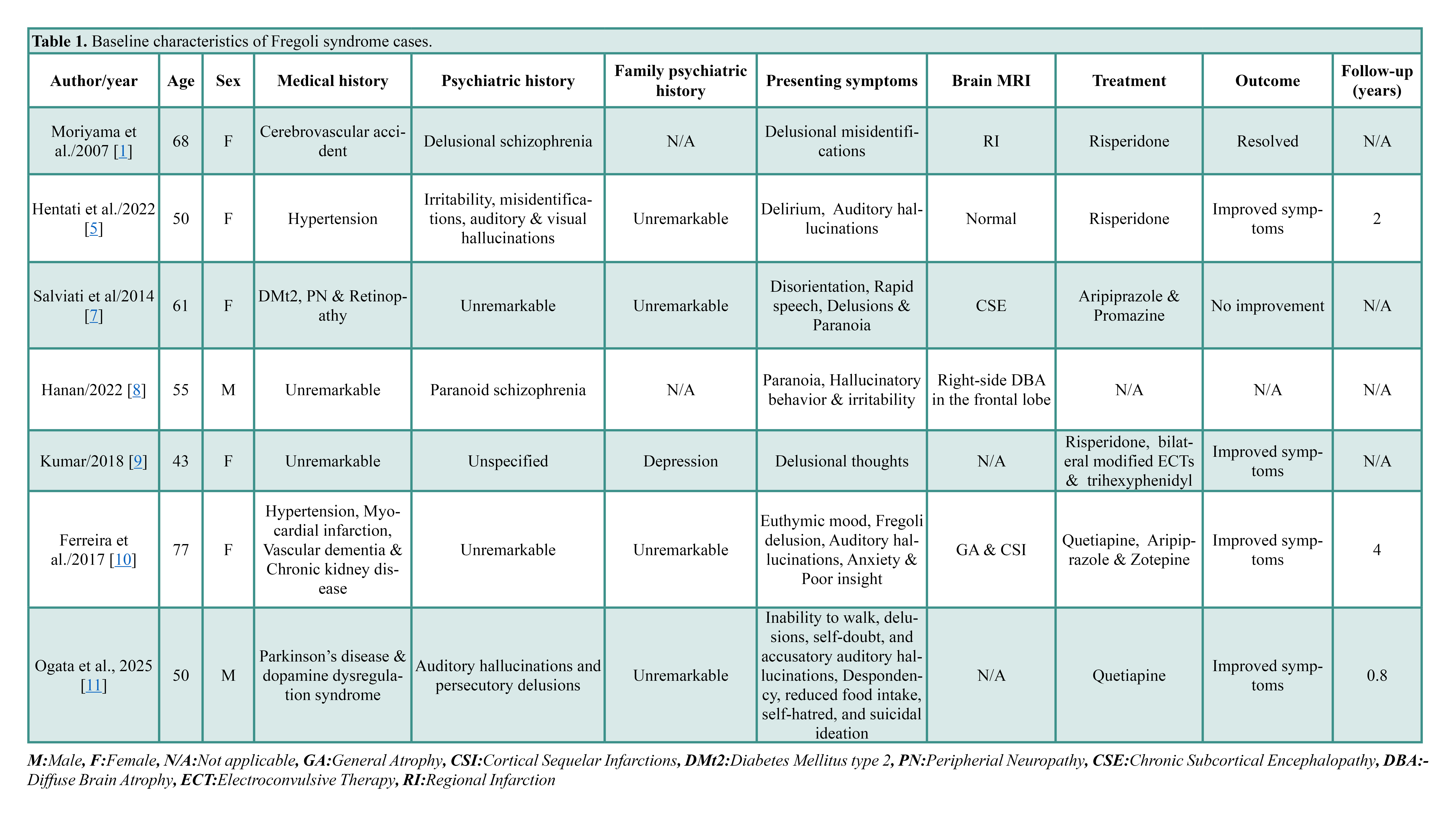
Literature review: Seven cases of Fregoli delusion were reviewed. Patient ages ranged from 43 to 77 years, with predominant female prevalence (71.4%). Psychiatric histories included delusional schizophrenia, paranoid schizophrenia, and unspecified psychiatric conditions. Medical histories included hypertension, myocardial infarction, Parkinson’s, and diabetes mellitus. Presenting symptoms varied with delusional misidentification, auditory/visual hallucinations, suicidal ideations, delirium, and paranoia. Treatment approaches included risperidone alone or in combination with electroconvulsive therapy, aripiprazole & promazine, and more. Symptom improvement was seen in four cases, and one case achieved full resolution of symptoms.
Conclusion: Fregoli syndrome is a rare condition of multiple etiologies that can present in young patients. Risperidone, combined with cognitive behavioral therapy, may yield fruitful results.
Hemn H. Kaka Ali, Abeer K. Abass, Ali H. Hasan, Ahmed H. Ahmed, Naser A. Mohammed, Twana O. Saeed, Marwan N. Hassan, Yadgar H. Hamakarim, Deari A. Ismaeil, Shvan H. Mohammed, Hussein M. Hamasalih, Fahmi H. Kakamad (Author)
Challenging Management of Postoperative Empyema: A Case Report with Literature Review
Hemn H. Kaka Ali, Abeer K. Abass, Ali H. Hasan, Ahmed H. Ahmed, Naser A. Mohammed, Twana O....
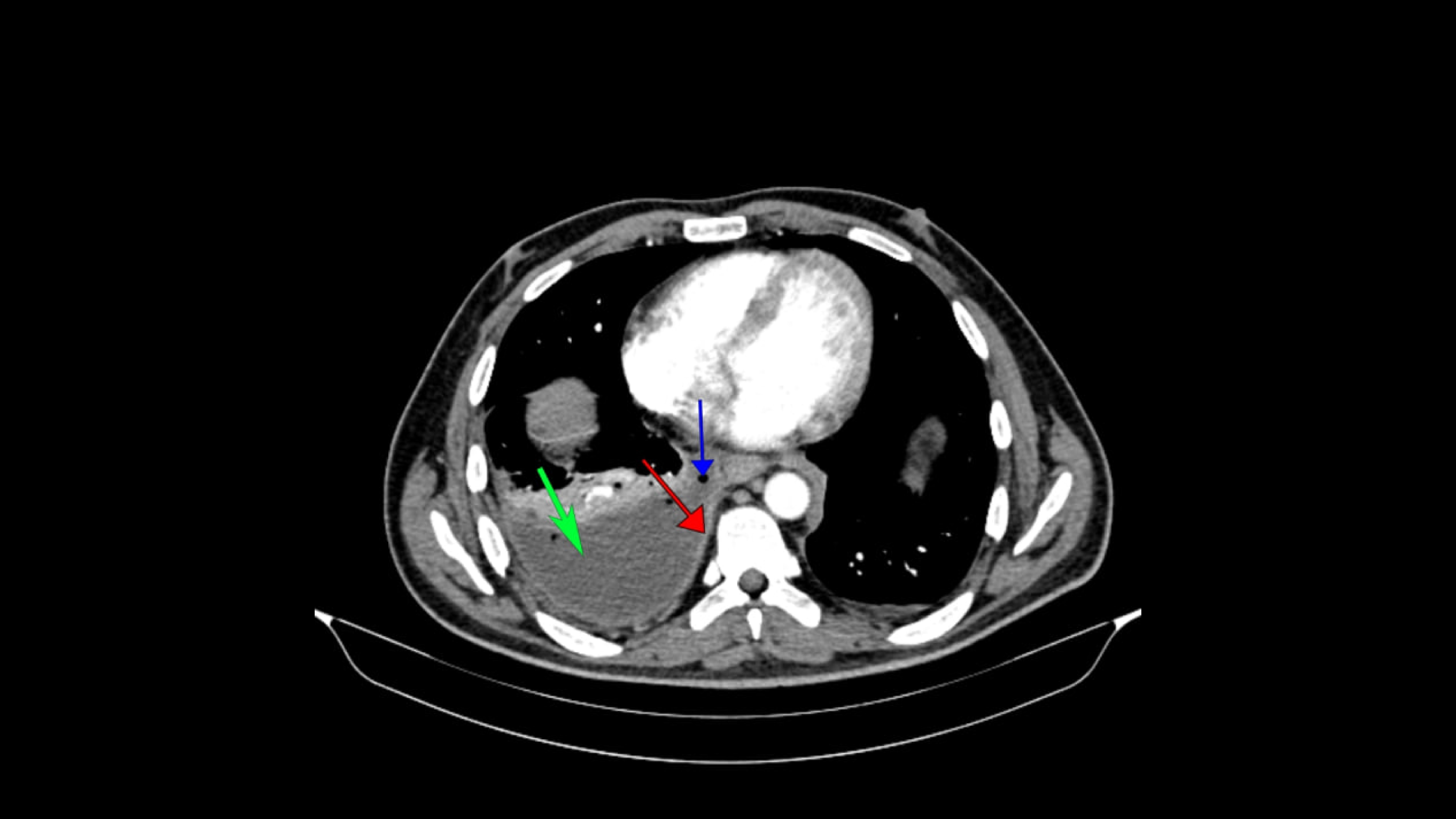
Introduction: Pleural empyema is the collection of pus within the pleural cavity, typically arising as a complication of pneumonia, chest trauma, thoracic surgery, or bacterial invasion of the pleural space. This report presents a case of post-surgical pleural empyema caused by Pseudomonas aeruginosa, successfully managed with a targeted combination of fosfomycin and colistin, with intrapleural lavage.
Case presentation: A 37-year-old male developed epigastric pain 12 days after a laparoscopic near-total gastrectomy. A chest computed tomography scan revealed a right-sided pleural empyema. Ultrasound-guided drainage was performed, followed by the intrapleural instillation of alteplase to facilitate breakdown of the loculated empyema. Pseudomonas aeruginosa was identified as the causative agent. Based on antimicrobial susceptibility, the patient received intravenous fosfomycin and colistin, along with daily pleural lavage using colistin. Inflammatory markers declined, and the patient showed notable clinical improvement.
Literature review: A review of five cases of Pseudomonas aeruginosa pleural empyema was conducted, including two carbapenem-resistant and one extensively drug-resistant case. The mean patient age was 53.8 years, and 60% (3/5) were female. Four of the five cases (80%) were confirmed using computed tomography, and all patients received antimicrobial therapy, most frequently ceftolozane/tazobactam (60%), ciprofloxacin (60%), and colistin (40%). Surgical management was required in 60% of cases, whereas bacteriophage therapy was utilized in 20%. During follow-up, 60% of patients remained stable, 20% experienced repeated hospital admissions during which antibacterial therapy was withheld, and 20% died due to infectious disease.
Conclusion: Pleural lavage combined with antibiotics such as fosfomycin and colistin may provide an effective treatment for postoperative pleural empyema, with early intervention being critical to prevent clinical deterioration.