Abdulwahid M. Salih, Ari M. Abdullah, Aso N. Qadir, Abdullah A. Qadir, Shko H. Hassan, Shalaw A. Nasraldeen, Saeed H. Ali, Muhammad H. Ali, Mohammed L. Ahmed, Osama A. Ali, Abdullah O. Hassan, Mohammed H. Rifaat, Daroo A. Kareem, Hawzhan Abubakr Abdalla, Sarkrda Mohammed Jamal (Author)
Abdulwahid M. Salih, Ari M. Abdullah, Aso N. Qadir, Abdullah A. Qadir, Shko H. Hassan, Shalaw A....
Introduction: Dyshormonogenetic goiter (DG) is a rare inherited cause of congenital hypothyroidism. Although DG is intrinsically benign, chronic thyroid-stimulating hormone stimulation has been implicated as a potential tumorigenic factor. This study is a single-center experience of the clinicopathological characteristics of DG.
Methods: The hospital registry database was filtered from February 2020 to December 2025 for cases of DG. The extracted data included clinical presentation, biochemical tests, imaging findings, diagnostic approaches, management, and outcomes. Only cases with clinicopathological findings consistent with DG were included.
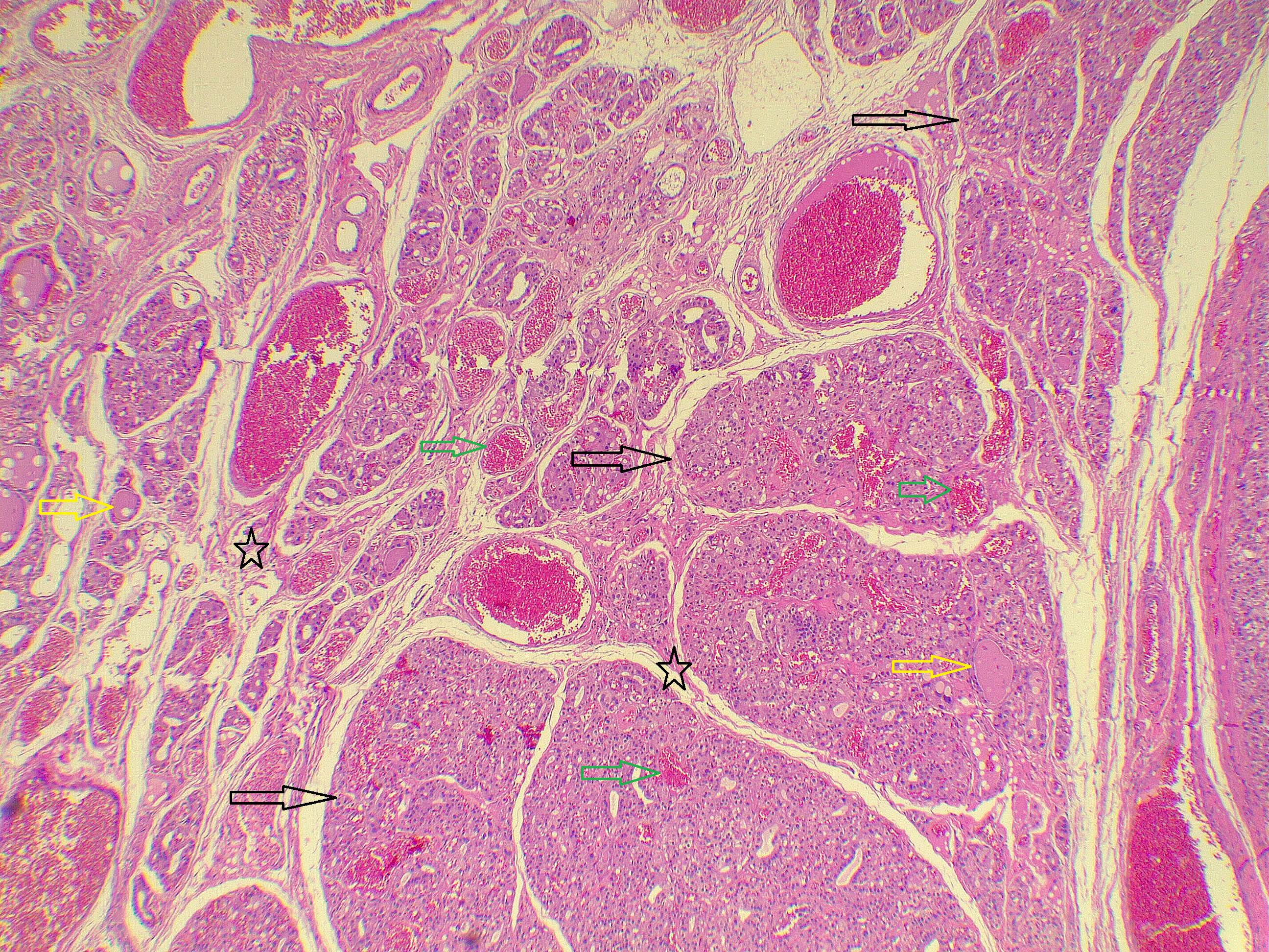
Results: The cohort included 15 patients (10 females, 5 males) with a median age of 17 years, ranging from 12-42 years. The disease was detected incidentally in 40.0% of cases (6/15). In total, 60.0% (9/15) had a history of congenital hypothyroidism or hypothyroidism. Ultrasonography demonstrated multinodular goiter in 93.3% (14/15), with high-risk features (TI-RADS 4 or 5) identified in 26.7% (4/15). Fine-needle aspiration cytology was performed in 4 patients, yielding Bethesda IV results in 3 cases and Bethesda II in 1 case. Histopathological examination revealed malignant neoplastic transformation in 20.0% (3/15) of cases, comprising two cases of follicular thyroid carcinomas (13.3%) and one papillary thyroid microcarcinoma (6.7%). On follow-up, all patients, except one, were euthyroid on levothyroxine.
Conclusion: Dyshormonogenetic goiter may closely mimic thyroid malignancy, and malignant transformation is also possible, which may support a role for chronic TSH stimulation as a tumorigenic stimulus, but larger multicenter studies with genetic characterization and longer follow-up are needed to define the true malignant potential of this entity.
Fahmi H. Kakamad, Shakew S. Mohammed, Razan Babarasul Jalal, Azad S. Hattam, Saywan K. Asaad, Abdullah K. Ghafour, Lawand A. Sharif, Huzaifa Hamid Ahmed, Hadeel Adnan Yasseen, Miran I. Hassan, Hiwa S. Namiq, Harun Amanj Ahmed (Author)
Vertebral Mass Mimicking Thoracic Outlet Syndrome: A Case Report with Literature Review
Fahmi H. Kakamad, Shakew S. Mohammed, Razan Babarasul Jalal, Azad S. Hattam, Saywan K. Asaad,...
Introduction: Neurogenic thoracic outlet syndrome is the most common subtype of thoracic outlet syndrome and can be readily mimicked and misdiagnosed. This report presents a rare cervical vertebral lesion mimicking neurogenic thoracic outlet syndrome, highlighting an important diagnostic pitfall.
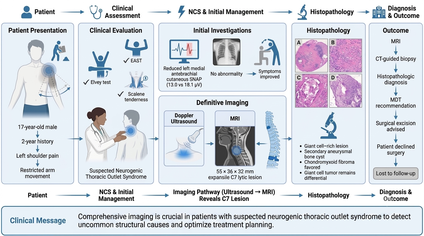
Case Presentation: Presenting with two years of left shoulder pain and immobility following heavy labor, a 17-year-old male underwent provocative testing and nerve conduction studies suggesting left neurogenic thoracic outlet syndrome. Despite improvement with physiotherapy, imaging revealed an expansile lytic mass at the C7 level, measuring 55 × 36 × 32 mm. Biopsy favored chondromyxoid fibroma. Surgical excision was decided, and the patient was lost to follow-up.
Literature Review: Five cases of cervicothoracic lesions presenting as thoracic outlet syndrome were reviewed, three of which were female. Pain and paresthesia were the most common symptoms, and the neurogenic subtype predominated. Most lesions arose from the first rib, and histology varied, including aneurysmal bone cyst, osteoblastoma, chondrosarcoma, and hydatid cyst. All patients underwent surgical excision, with symptom resolution or improvement.
Conclusion: Cervical vertebral lesions can mimic neurogenic thoracic outlet syndrome, highlighting the importance of imaging in identifying underlying structural pathology.