
Dyshormonogenetic Goiter: A Case Series

Abstract
Introduction: Dyshormonogenetic goiter (DG) is a rare inherited cause of congenital hypothyroidism. Although DG is intrinsically benign, chronic thyroid-stimulating hormone stimulation has been implicated as a potential tumorigenic factor. This study is a single-center experience of the clinicopathological characteristics of DG.
Methods: The hospital registry database was filtered from February 2020 to December 2025 for cases of DG. The extracted data included clinical presentation, biochemical tests, imaging findings, diagnostic approaches, management, and outcomes. Only cases with clinicopathological findings consistent with DG were included.
Results: The cohort included 15 patients (10 females, 5 males) with a median age of 17 years, ranging from 12-42 years. The disease was detected incidentally in 40.0% of cases (6/15). In total, 60.0% (9/15) had a history of congenital hypothyroidism or hypothyroidism. Ultrasonography demonstrated multinodular goiter in 93.3% (14/15), with high-risk features (TI-RADS 4 or 5) identified in 26.7% (4/15). Fine-needle aspiration cytology was performed in 4 patients, yielding Bethesda IV results in 3 cases and Bethesda II in 1 case. Histopathological examination revealed malignant neoplastic transformation in 20.0% (3/15) of cases, comprising two cases of follicular thyroid carcinomas (13.3%) and one papillary thyroid microcarcinoma (6.7%). On follow-up, all patients, except one, were euthyroid on levothyroxine.
Conclusion: Dyshormonogenetic goiter may closely mimic thyroid malignancy, and malignant transformation is also possible, which may support a role for chronic TSH stimulation as a tumorigenic stimulus, but larger multicenter studies with genetic characterization and longer follow-up are needed to define the true malignant potential of this entity.
Introduction
Congenital hypothyroidism (CH) is the most common neonatal endocrine disorder, defined as thyroid hormone insufficiency at birth. Etiologically, primary CH comprises thyroid dysgenesis, reflecting aberrant glandular development, and thyroid dyshormonogenesis, denoting inherited defects of hormone biosynthesis within a structurally formed gland. The term dyshormonogenetic goiter (DG), by contrast, designates the morphological sequela of these defects [1,2]. Dyshormonogenetic goiter is a rare clinical entity first documented in the late 19th century by Pendred and Osler [3]. Unlike dysgenesis, which involves glandular agenesis or ectopia, dyshormonogenesis is characterized by an anatomically orthotopic gland. In these cases, congenital metabolic errors impede hormone biosynthesis, leading to chronic thyroid-stimulating hormone (TSH) stimulation that drives compensatory hyperplasia, goiter formation, and significant architectural distortion [4-6]. These features often mimic thyroid malignancy, creating substantial diagnostic challenges and a risk of overdiagnosis [3,4]. While the incidence of primary CH has risen to approximately 1 in 2,000 to 1 in 3,000 newborns, DG remains rare, with prevalence estimates ranging between 1 in 30,000 and 1 in 50,000 live births [1,3]. It is a genetic condition, typically inherited in an autosomal recessive pattern, arising from molecular aberrations in key proteins required for thyroid hormone biosynthesis [5,7-9]. Although DG is intrinsically benign, chronic TSH stimulation has been implicated as a potential tumorigenic factor [10-12]. This study is a single-center experience of the clinicopathological characteristics of DG.
Methods
Study Design and Setting
This study was conducted in the Head and Neck Surgery Department at Smart Health Tower. The data were collected retrospectively from the department registry database for all cases that sought consultation for thyroid-related complaints from February 2020 to December 2025.
Participant Selection and Eligibility Criteria
The study eligibility criteria were limited to patients with clinicopathological findings consistent with DG (elevated TSH from childhood, orthotopic gland, characteristic histology, absence of autoimmunity by anti-TPO, family history) and with complete medical records. Therefore, cases with other thyroid abnormalities except those that were concomitant with DG, and incomplete medical records (cases with no reported clinical and pathological data) were excluded. No age, sex, or ethnicity restrictions were applied in the data retrieval.
Data Collection
Patient data were extracted from electronic medical records, radiological archives, and clinical documentation. The data included clinical presentation, biochemical tests, imaging findings, diagnostic approaches, management, and outcomes.
Data Analysis
Data entry and descriptive analysis were conducted using Microsoft Excel 2016. The distribution of the continuous variable (age) was assessed by the Shapiro-Wilk test using IBM SPSS (version 27). The variable was non-normal, so it was reported as median (quartile ranges) and range. The qualitative variables were reported as frequencies and percentages.
Results
This study included 15 patients with a median age of 17 years (range: 12-42 years). The cohort comprised 10 females (66.7%) and 5 males (33.3%). Two patients (Cases 4 and 5) were siblings. Six patients (40.0%) were diagnosed incidentally during routine follow-up for congenital hypothyroidism or after an incidental ultrasonography (U/S) finding, while 8 (53.3%) presented with anterior neck swelling, and 1 (6.7%) presented with facial swelling accompanied by tongue protrusion. A history of CH or hypothyroidism was documented in 9 cases (60.0%), bronchial asthma in one (6.7%), and no significant past medical history in 5 (33.3%). A family history of thyroid disease was present in 4 patients (26.7%). Goiter grading based on WHO classification [13] showed G1 in 9 cases (60.0%), G2 in 4 cases (26.7%), G3 in 1 case (6.7%), and G0 in 1 case (6.7%) (Table 1).
|
Case No. |
Age (Y) |
Gender |
History |
Clinical Thyroid Examinations |
|||||
|
Presentation |
PMH |
PSH |
FHX |
Medication Hx |
Smoking |
||||
|
Case 1 |
17 |
M |
Thyroid check-up |
Congenital Hypothyroidism |
None |
None |
T4 100 μg |
No |
G0 |
|
Case 2 |
16 |
F |
Neck swelling |
Congenital Hypothyroidism |
None |
Thyroid disease, DM, HTN |
T4 100 μg |
No |
G2 |
|
Case 3 |
16 |
M |
Neck swelling |
Congenital Hypothyroidism |
None |
None |
T4 75 μg |
No |
G2 |
|
Case 4 |
12 |
F |
Thyroid check-up |
Congenital Hypothyroidism |
None |
Dyshormonogenesis, Papillary Thyroid Carcinoma |
T4 75 μg |
No |
G1 |
|
Case 5 |
15 |
F |
Thyroid check-up |
Bronchial Asthma |
None |
Dyshormonogenesis, Papillary Thyroid Carcinoma |
None |
No |
G1 |
|
Case 6 |
32 |
F |
Thyroid check-up |
None |
None |
None |
None |
No |
G3 |
|
Case 7 |
26 |
M |
Thyroid check-up |
None |
None |
None |
None |
No |
G1 |
|
Case 8 |
21 |
F |
Neck swelling |
None |
None |
None |
None |
No |
G2 |
|
Case 9 |
21 |
F |
Facial swelling, Tongue protrusion |
None |
None |
None |
None |
No |
G2 |
|
Case 10 |
17 |
F |
Neck swelling |
None |
None |
None |
None |
No |
G1 |
|
Case 11 |
16 |
M |
Thyroid check-up |
Congenital Hypothyroidism |
None |
None |
T4 100 μg |
No |
G1 |
|
Case 12 |
18 |
M |
Neck swelling |
Congenital Hypothyroidism |
Eye surgery |
Hypothyroidism |
T4 100 μg |
No |
G1 |
|
Case 13 |
14 |
F |
Neck swelling |
Congenital Hypothyroidism |
None |
None |
T4 100 μg |
No |
G1 |
|
Case 14 |
32 |
F |
Neck swelling |
Hypothyroidism |
None |
None |
T4 75 μg |
No |
G1 |
|
Case 15 |
42 |
F |
Neck swelling |
Hypothyroidism |
None |
None |
T4 75 μg |
No |
G1 |
|
Abbreviations. No., Number; Y, Year; M, Male; F, Female; PMH, Past Medical History; PSH, Past Surgical History; FHX, Family History; DM, Diabetes Mellitus; HTN, Hypertension; Hx, History; T4, Levothyroxine; µg, Microgram; G, Grade (of Goiter). |
|||||||||
Preoperative biochemical assessment showed elevated TSH across the cohort, except one case (was on levothyroxine due to congenital hypothyroidism), ranging from 5.96 to >100 mIU/L (reference range: 0.35-4.94 mIU/L), with fT4 values spanning 0.55-20.63 pmol/L (reference range: 9.0-19.0 pmol/L) and Anti-TPO levels between 5.00 and 18.46 IU/mL (reference range: <34 IU/mL). Ultrasonography demonstrated multinodular goiter in 14 patients (93.3%) and a solitary nodule in 1 (6.7%), with the Thyroid Imaging Reporting and Data System (TI-RADS) category recorded as TI-RADS 2 in 1 case (6.7%), TI-RADS 3 in 11 (73.3%), TI-RADS 4 in 4 (26.7%), and TI-RADS 5 in 1 (6.7%). TI-RADS was not specified in two cases (13.3%). The largest documented thyroid lobe was the right lobe in case 6, measuring 120 × 53 × 51 mm. Ultrasound-guided fine needle aspiration cytology was performed in 4 patients, yielding Bethesda IV results in 3 cases and Bethesda II in 1 case (Table 2).
|
Case No. |
Biochemical Profile |
Neck Ultrasound |
FNAC |
Surgical Procedure |
HPE |
Postoperative Management |
Postoperative TSH (mIU/L) |
Follow-Up Status |
Post-operative follow-up duration (years) |
|||||
|
TSH (mIU/L) |
fT4 (pmol/L) |
Anti-TPO (IU/mL) |
Findings |
Right Lobe Size (mm) |
Left Lobe Size (mm) |
Nodule Characteristics |
||||||||
|
Case 1 |
67.59 |
10.65 |
7.89 |
Thyroiditis |
65 × 20 × 18 |
60 × 20 × 18 |
Multinodular Goiter; 14 × 7 mm nodule at the inferior junction of the Right Lobe and isthmus. |
Not performed |
Total Thyroidectomy |
DG with multiple adenomatoid nodules |
T4 100 µg |
3 |
Alive, Euthyroid |
1 |
|
Case 2 |
100.00 |
0.55 |
5.00 |
Thyroiditis |
40 × 13 × 12 |
37 × 13 × 12 |
Multinodular Goiter. Right Lobe: 18 × 8 mm well-defined hypoechoic nodule with punctate echogenic foci (ACR TI-RADS 5); 8 × 4 mm (ACR TI-RADS 4); 2 × 2 mm well-defined hypoechoic nodule (ACR TI-RADS 4). Left Lobe: 11 × 8 mm nodule with perinodular vascularity but no intra nodular vascularity (ACR TI-RADS 4). |
Not performed |
Total Thyroidectomy |
DG with multiple adenomatoid nodules |
T4 100 µg |
0.9 |
Alive, Euthyroid |
3 |
|
Case 3 |
>100.00 |
4.36 |
6.54 |
Thyroiditis |
77 × 33 × 32 |
70 × 26 × 21 |
Multinodular Goiter. Right Lobe: Well-defined, predominantly solid, iso- to hyperechoic nodule (43 × 30 ×25 mm) in upper-mid third (ACR TI-RADS 3); two similar nodules (14 × 13 × 12 mm in mid-third; 21 × 14 × 12 mm in lower-third). Left Lobe: Two small nodules, largest 5 × 3 mm (ACR TI-RADS 2). |
Not performed |
Total Thyroidectomy |
DG |
T4 100 µg |
4.05 |
Alive, Euthyroid |
1 |
|
Case 4 |
41.59 |
9.75 |
12.22 |
Thyroiditis |
65 × 23 × 20 |
60 × 22 × 20 |
Multinodular Goiter. Right Lobe: 20 × 16 × 14 |
Not performed |
Total Thyroidectomy |
DG |
T4 100 µg |
4.37 |
Alive, Euthyroid |
4 |
|
Case 5 |
20.89 |
12.08 |
11.11 |
Multinodular Goiter |
65 × 15 × 14 |
60 × 13 × 13 |
Multinodular Goiter. Right Lobe: 11 × 8 × 6 mm nodule (ACR TI-RADS 3). Left Lobe: Largest nodule 11 × 7 × 5 mm (ACR TI-RADS 3). |
Not performed |
Total Thyroidectomy |
DG with multiple adenomatoid nodules |
T4 100 µg |
0.43 |
Alive, Euthyroid |
2 |
|
Case 6 |
10.67 |
7.65 |
18.46 |
Multinodular Goiter |
120 × 53 × 51 |
110 × 43 × 35 |
Multinodular Goiter. Right Lobe: 40 × 32 × 30 mm (ACR TI-RADS 3) and 29 × 20 × 18 mm (ACR TI-RADS 4). Left Lobe: Largest nodule 22 × 18 × 17 mm (ACR TI-RADS 3) and 16 × 15 × 12 mm (ACR TI-RADS 4). |
Bethesda IV |
Total Thyroidectomy |
Follicular Thyroid Carcinoma with Complete capsular and vascular (at least two vessels) invasion arising from DG |
T4 100 µg |
5.74 |
Alive, No Recurrence with slightly elevated TSH |
3 |
|
Case 7 |
100.00 |
2.52 |
13.10 |
Multinodular Goiter |
50 × 17 × 15 |
65 × 44 × 34 |
Multinodular Goiter. Right Lobe: 10 × 6 × 5 mm nodule (ACR TI-RADS 4). Left Lobe: Large (57 × 41 × 29 mm), well-defined, regular, predominantly solid nodule with macrocalcification and increased perinodular/intranodular vascularity (ACR TI-RADS 4). |
Bethesda IV |
Total Thyroidectomy |
Follicular Thyroid Carcinoma with Complete capsular and vascular (at least 6 vessels) invasion arising from DG |
T4 100 µg |
3.26 |
Alive, Euthyroid |
1 |
|
Case 8 |
53.25 |
9.75 |
7.20 |
Thyroiditis |
60 × 32 × 25 |
43 × 14 × 14 |
Solitary Nodule. Well-defined, regular surface, solid nodule (37 × 29 × 23 mm) occupying most of the gland. |
Bethesda IV |
Total Thyroidectomy |
Follicular Adenoma arising from DG |
T4 150 µg |
0.91 |
Alive, Euthyroid |
1 |
|
Case 9 |
47.41 |
8.30 |
5.40 |
Thyroiditis |
70 × 26 × 24 |
80 × 27 × 25 |
Multinodular Goiter. Right Lobe: Largest nodule 19 × 14 mm (ACR TI-RADS 3). Left Lobe: Largest nodule 31 × 22 mm (ACR TI-RADS 3). |
Not performed |
Total Thyroidectomy |
DG with multiple adenomatoid nodules |
T4 150 µg |
4.43 |
Alive, Euthyroid |
3 |
|
Case 10 |
5.96 |
17.86 |
9.13 |
Multinodular Goiter |
77 × 40 × 37 |
80 × 42 × 38 |
Multinodular Goiter. Right Lobe: Largest nodule 36 × 31 × 28 mm (ACR TI-RADS 3). Left Lobe: Largest nodule 29 × 25 mm (ACR TI-RADS 3). |
Not performed |
Total Thyroidectomy |
DG with multiple adenomatoid nodules |
T4 100 µg |
3.39 |
Alive, Euthyroid |
2 |
|
Case 11 |
0.67 |
20.63 |
5.40 |
Multinodular Goiter |
95 × 45 × 40 |
77 × 41 × 33 |
Multinodular Goiter. Right Lobe: Largest nodule 32 × 25 × 24 mm (ACR TI-RADS 3). Left Lobe: Largest nodule 25 × 22 mm (ACR TI-RADS 3). |
Not performed |
Total Thyroidectomy |
DG with multiple adenomatoid nodules |
T4 100 µg |
4.39 |
Alive, Euthyroid |
2 |
|
Case 12 |
16.06 |
20.01 |
5.09 |
Multinodular Goiter |
60 × 24 × 21 |
56 × 20 × 18 |
Multinodular Goiter. Right Lobe: Largest nodule 22 × 16 × 14 mm (ACR TI-RADS 3). Left Lobe: Largest nodule 18 × 10 mm (ACR TI-RADS 3). |
Bethesda II |
Total Thyroidectomy |
Papillary Thyroid Microcarcinoma, Conventional Subtype (0.5 cm), Unifocal, Left Thyroid Lobe, without Extrathyroidal Extension, Lymphovascular or Perineural Invasion, arising in DG with Multiple Bilateral Adenomatoid Nodules (pT1aN0). |
T4 100 µg |
4.59 |
Alive, Euthyroid |
1 |
|
Case 13 |
5.96 |
17.86 |
7.13 |
Multinodular Goiter |
80 × 41 × 36 |
100 × 60 × 44 |
Multinodular Goiter. Right lobe: Largest nodule 40 × 35 × 27 mm (ACR TI-RADS 3). Left Lobe: Largest nodule 36 × 28 mm (ACR TI-RADS 3). |
Not performed |
Total Thyroidectomy |
DG with multiple adenomatoid nodules |
T4 100 µg |
2.59 |
Alive, Euthyroid |
4 |
|
Case 14 |
>100 |
4.59 |
15.60 |
Multinodular Goiter |
67 × 32 × 29 |
60 × 29 × 27 |
Multinodular Goiter. Right Lobe: Largest nodule 34 × 30 × 23 mm (ACR TI-RADS 3). Left Lobe: Largest nodule 26 × 20 × 13 mm (ACR TI-RADS 3). |
Not performed |
Total Thyroidectomy |
DG with multiple adenomatoid nodules |
T4 100 µg |
2.59 |
Alive, Euthyroid |
3 |
|
Case 15 |
14.05 |
7.99 |
10.02 |
Multinodular Goiter |
80 × 44 × 35 |
70 × 30 × 24 |
Multinodular Goiter. Right Lobe: Largest nodule 39 × 23 × 20 mm (ACR TI-RADS 3). Left Lobe: Largest nodule 14 × 10 × 8 mm (ACR TI-RADS 4). |
Not performed |
Total Thyroidectomy |
DG with multiple adenomatoid nodules |
T4 100 µg |
1.09 |
Alive, Euthyroid |
2 |
|
Abbreviations. No., Number; TSH, Thyroid Stimulating Hormone; mIU/L, Milli-international units per liter; fT4, Free Thyroxine; pmol/L, Picomoles per liter; Anti-TPO, Anti-thyroid Peroxidase Antibody; IU/mL, International units per milliliter; mm, Millimeter; ACR, American College of Radiology; TI-RADS, Thyroid Imaging Reporting and Data System; FNAC, Fine Needle Aspiration Cytology; Bethesda IV, Suspicious for Follicular Neoplasm; HPE, Histopathological Examination; Bethesda II, Benign. DG, Dyshormonogenetic Goiter; µg, Microgram; T4, Levothyroxine. |
||||||||||||||
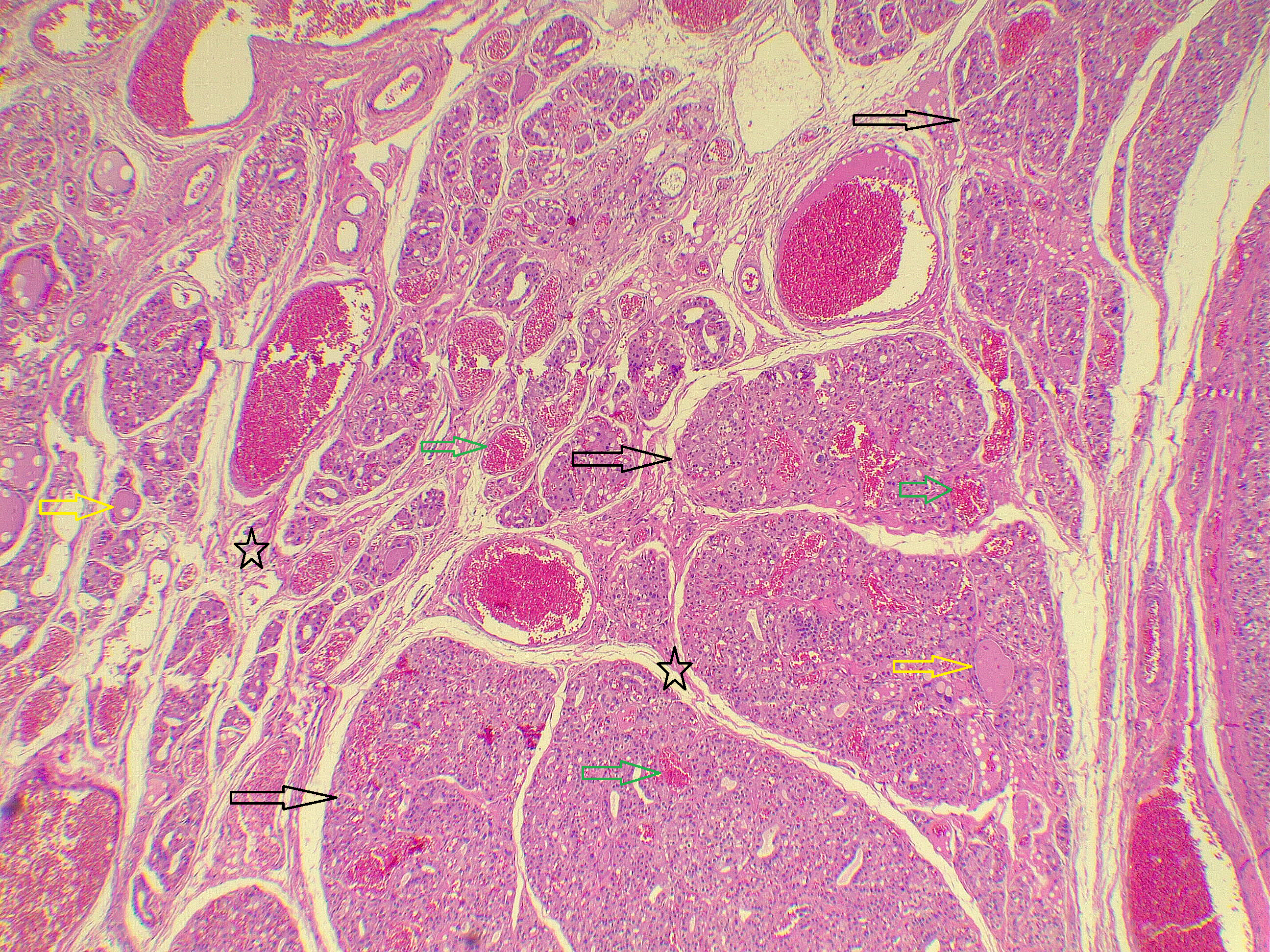
All patients underwent total thyroidectomy, and the rationale included high goiter grading (cases 3,6,8,9) and congenital hypothyroidism with compressive symptoms/inability to exclude malignancy (cases 1-4,11-13), TI-RADS category > 3 (cases 2,6,7,15), Bethesda ≥ IV (cases 6 to 8), and nodular sizes greater than 2.5 cm on U/S (cases 3,6-11,13-15) and positive family history for thyroid cancer and dyshormonogenesis (cases 4,5). Histopathological examination revealed malignant neoplastic transformation in 20.0% (3/15) of cases, comprising two cases of follicular thyroid carcinomas (13.3%) and one papillary thyroid microcarcinoma (6.7%). On microscopic examination, sections showed irregularly sized thyroid follicles with scant colloid and areas of empty lumina, with background fibrosis and multiple adenomatoid nodules, and focal marked nuclear atypia (Figure 1). The two patients with follicular thyroid carcinoma underwent radioactive iodine therapy, and the other case of papillary thyroid microcarcinoma remained under regular follow-up. Postoperatively, all patients were alive and clinically stable on levothyroxine doses of 100-150 µg daily. TSH values, 8 weeks after starting replacement therapy, were within the reference range for 14 patients (0.43-4.59 mIU/L), while one patient had a slightly elevated level of 5.74 mIU/L (reference range: 0.35-4.94 mIU/L). The follow-up period ranged from 1 year to 4 years (Table 2 & 3).
|
Variable |
Frequency (n)/Median (Q1-Q3)/Range |
Percentage (%) |
|
Gender Male Female |
5 10 |
33.3% 66.7% |
|
Age (Yrs) Median (Q1-Q3) Range |
17 (16-26) 12-42 |
|
|
Clinical Presentation Incidental finding Neck swelling Facial swelling/Tongue protrusion
|
6 8 1
|
40.0% 53.3% 6.7%
|
|
Past Medical History Congenital Hypo/Hypo Bronchial Asthma No significant history |
9 1 5 |
60.0% 6.7% 33.3%
|
|
Clinical Thyroid Examination G 0 G 1 G 2 G 3 |
2 9 3 1 |
13.3% 60.0% 20.0% 6.7%
|
|
Family History of Thyroid Disease Positive Negative |
4 11 |
26.7% 73.3% |
|
Ultrasound Features Multinodular Goiter Solitary nodule |
14 1 |
93.3% 6.7% |
|
ACR TI-RADS Score Per Case* TI-RADS 2 TI-RADS 3 TI-RADS 4 TI-RADS 5 Not specified |
1 11 4 1 2 |
6.7% 73.3% 26.7% 6.7% 13.3%
|
|
Other pathologies besides DG Multiple adenomatoid nodules Follicular Thyroid Carcinoma Papillary thyroid microcarcinoma Follicular Adenoma None |
10 2 1 1 2 |
66.7% 13.3% 6.7% 6.7% 13.3%
|
|
*Each case may have lesions with different TI-RADS Abbreviations. N, (number of patients in a specific category); Yrs, Years; G, Grade (of goiter); ACR, American College of Radiology; TI-RADS, Thyroid Imaging Reporting and Data System; Hypo, Hypothyroidism; DG, Dyshormonogenetic Goiter. |
||
Discussion
The clinical presentation of DG is classically documented as a neonatal or infantile condition, often manifesting immediately after birth. Previous reports have documented neonatal presentations with clinically significant airway compression, including a case involving a 4-day-old infant, as well as large cervical masses detected as early as 11 hours after birth [2,14]. Additionally, fetal goiters may cause complications such as polyhydramnios in utero [2]. In contrast to these findings, the diagnosis in this series was established at a median age of 17 years in accordance with the clinicopathological study by Ghossein et al., which reported a median age of approximately 16 years at surgery [15]. This delayed presentation aligns with reports of adolescents and young adults who remain undiagnosed until metabolic demands peak, as in cases of a 19-year-old male and a 10.5-year-old boy recently reported [7,16]. This spectrum of early-onset disease includes genetically defined cases, such as siblings with fetal goiters caused by compound heterozygous TPO gene mutations, which can also affect long-term neuropsychological outcomes [17]. The exclusivity of adolescent and adult patients in this case series (≥12 years old) corroborates observations from other resource-limited settings, where diagnosis is frequently delayed by over a decade, supporting the hypothesis of "compensated dyshormonogenesis", which permits subclinical progression into adulthood [4].
The clinical presentation observed in this study differs markedly from the acute manifestations seen in neonates. While several reported neonatal cases present with life-threatening stridor or severe neck extension [4,8,15], the clinical picture observed in the present study was predominantly insidious. Overall, 40.0% of the patients were identified incidentally, a finding that contrasts with case reports from India, where older children consistently presented with overt neck swelling and mechanical symptoms like dyspnea [3,4]. However, this high rate of incidental detection aligns with a study, where siblings of index patients were found to have goiters despite being clinically asymptomatic [10]. The absence of acute symptoms in a subset of the present cases, despite significant glandular enlargement in some, reinforces the insidious nature of partial enzymatic defects, which can allow patients to remain clinically compensated for years before structural changes necessitate evaluation [7].
Radiological evaluation often reveals features overlapping significantly with thyroid malignancy, complicating risk stratification. Literature indicates that benign dyshormonogenetic nodules frequently exhibit solid composition and hypoechogenicity, characteristics traditionally associated with malignancy, often leading to high TI-RADS scores [6]. In this study, U/S identified several nodules with high-risk features, which were classified as TI-RADS 4 or 5. This rate of suspicious findings is similar to pediatric cases and supports the decision for total thyroidectomy to rule out cancer [4]. Although benign features like a hypoechoic "halo" rim have been reported, the heterogeneous presentation underscores the diagnostic limitations of U/S in DG, where chronic TSH-driven hyperplasia creates architectural distortions that mimic neoplastic processes [6].
Histopathological evaluation of DG poses significant diagnostic challenges due to the profound architectural and cellular pleomorphism induced by chronic TSH stimulation. Characteristic features such as hypercellularity, nuclear atypia, and scant colloid, as described in the literature, can easily lead to a misdiagnosis of follicular or papillary thyroid carcinoma [3]. Furthermore, the presence of mitotically active follicular nodules has been reported to raise suspicion for poorly differentiated carcinoma, necessitating careful differentiation [18]. In the present study, histological confirmation of DG displayed irregular thyroid follicles with scant colloid, extensive background fibrosis, and multiple adenomatoid nodules. These findings align with observations that chronic TSH stimulation leads to a spectrum of proliferative changes, ranging from benign hyperplasia to lesions with uncertain malignant potential [6].
Beyond diagnostic mimicry, DG carries a potential risk of carcinogenesis. Although intrinsically benign, the chronic, high-intensity TSH stimulation serves as a potent proliferative driver that may promote mutagenesis over time. This risk is further amplified in patients with coexisting genetic syndromes; for instance, the combination of a TPO mutation with a PTEN mutation in Bannayan-Riley-Ruvalcaba syndrome may play a synergistic role in the development of multinodular goiter and increase the risk of malignant transformation [19]. Although rare, associations exist between dyshormonogenesis and the development of malignancies, including papillary thyroid carcinoma with lung metastasis and follicular thyroid carcinoma in patients with TPO mutations [20,21]. More aggressive outcomes, such as anaplastic thyroid cancer, have also been reported in association with specific TG gene mutations [11]. In this series, a malignant neoplastic transformation rate was observed, with 20.0% of patients diagnosed with thyroid cancer, including two cases of follicular thyroid carcinoma and a case of papillary thyroid microcarcinoma. This incidence may support the hypothesis that prolonged TSH elevation, potentially coupled with specific genetic backgrounds such as germline DUOX2 mutations, contributes to tumor progression [12].
The genetic landscape of thyroid dyshormonogenesis is highly heterogeneous, involving pathogenic variants in genes encoding key enzymes and transporters. While TPO mutations are traditionally classified as a primary cause of total iodide organification defects, recent large-scale cohorts have elucidated DUOX2 variants as a frequent etiology associated with a broad phenotypic spectrum [1,5,9]. Additionally, specific TG gene mutations causing intracellular transport defects can lead to goiter formation even with normal TSH levels [22]. Although molecular genetic testing was not performed in this series, a positive family history of thyroid disease in 26.7% of cases and having 2 sibling cases may be consistent with the predominantly autosomal recessive inheritance pattern typical of these metabolic disorders [5]. The clinical and histopathological findings strongly suggest underlying defects in the hormone biosynthetic pathway similar to those described in genetically characterized cohorts [1,9].
Management strategies for DG are dictated by the age of presentation and the severity of glandular enlargement. While early levothyroxine replacement in neonates can successfully reduce goiter size and relieve airway compression, the structural alterations characterized by dense fibrosis and multiple adenomatoid nodules in older patients are likely irreversible with medical suppression alone [2,3,21]. Consequently, total thyroidectomy served as the definitive management for all patients in this series, addressing both compressive symptoms due to neck swelling and the need to rule out malignancy in suspicious nodules. This approach is supported by recent literature advocating for surgery when conservative management fails or when malignancy cannot be excluded [4]. Postoperative outcomes were favorable, with nearly all patients achieving a stable euthyroid status on replacement therapy. Despite the limitation regarding genetic analysis (too costly for patients), this study highlights the significant risk of neoplastic transformation in patients with delayed diagnosis, underscoring the necessity for vigilant surveillance and a lower threshold for surgical intervention [21]. Other limitations included the study design, the relatively short follow-up period, and the inability to retrieve histopathological images for cases associated with malignancy, which limited further characterization of the disease and may have reduced the generalizability of the findings.
Conclusion
Dyshormonogenetic goiter may closely mimic thyroid malignancy, and malignant transformation is also possible, which may support a role for chronic TSH stimulation as a tumorigenic stimulus, but larger multicenter studies with genetic characterization and longer follow-up are needed to define the true malignant potential of this entity.
Declarations
Conflicts of Interest: The authors have no conflicts of interest to disclose.
Ethical Approval: Not applicable.
Consent for Participation: Not applicable.
Consent for Publication: Informed consent for the publication of anonymized clinical data and images was obtained from all patients (or their legal guardians, where applicable).
Funding: The present study received no financial support.
Acknowledgements: None to be declared.
Authors' Contributions: AMS, AMA and ANQ: Major contributors to the conception of the study, as well as the literature search for related studies, and manuscript writing. AAQ, SHH, SAN, SHA, MHA, MLA, OAA, AOH, MHR, DAK, HAA, SMJ: Literature review, critical revision of the manuscript, and processing of the tables.
Use of AI: ChatGPT (GPT-5.5, OpenAI) was used to assist with language refinement and improve the overall clarity of the manuscript. All content was thoroughly reviewed and approved by the authors, who bear full responsibility for the final version.
Data Availability Statement: All data generated or analyzed during this study are included in this published article. Further enquiries can be directed to the corresponding author.
References
- Su Y, Lei X, Muhetaer A, He J, Li L. Correlation of genotype-phenotype of congenital hypothyroidism cohort diagnosed by newborn screening: a long-term observational study. Int J Neonatal Screen. 2025;11(4):98. doi:10.3390/ijns11040098
- Hirano Y, Shimura K, Mitani-Konno M, Fukushima N, Hasegawa T, Narumi S. Airway compression in a neonate with thyroid dyshormonogenesis due to SLC26A7 defect. Eur J Endocrinol. 2025;193(4):K16-21. doi:10.1093/ejendo/lvaf198
- Momin YA, Ansari SAH. Dyshormonongenetic goiter: a rare and important entity. Arch Med Health Sci. 2020;8(1):88-90. doi:10.4103/amhs.amhs_152_19
- Dekhne A, Popat A, Chopwad A. Looking beyond thyroid malignancy: an unusual case of dyshormonogenetic goiter in rural India. Cureus. 2024;16(8):e68139. doi:10.7759/cureus.68139
- Arteaga-Jacobo MC, Roco-Videla Á, Villota Arcos C, González-Hormazábal P, Gonzalo-Castro V, Pérez-Flores MV. Frequency of mutations in the TPO gene in patients with congenital hypothyroidism due to dyshormonogenesis in Chile. Medicina (Kaunas). 2024;60(7):1145. doi:10.3390/medicina60071145
- Januś D, Kujdowicz M, Kiszka-Wiłkojć A, Kaleta K, Taczanowska-Niemczuk A, Radliński J, et al. Ultrasound and histopathological assessment of benign, borderline, and malignant thyroid tumors in pediatric patients: an illustrative review and literature overview. Front Endocrinol (Lausanne). 2025;15:1481804. doi:10.3389/fendo.2024.1481804
- Sciarroni E, Montanelli L, Di Cosmo C, Bagattini B, Comi S, Pignata L, et al. Late-onset dyshormonogenic goitrous hypothyroidism due to a homozygous mutation of the SLC26A7 gene: a case report. Ital J Pediatr. 2024;50(1):106. doi:10.1186/s13052-024-01672-3
- Stern E, Schoenmakers N, Nicholas AK, Kassif E, Pinhas Hamiel O, Yeshayahu Y. A novel mutation in the thyroglobulin gene resulting in neonatal goiter and congenital hypothyroidism in an Eritrean infant. J Clin Res Pediatr Endocrinol. 2022;14(2):221-6. doi:10.4274/jcrpe.galenos.2021.2020.0278
- Baz-Redón N, Antolín M, Clemente M, Campos A, Mogas E, Fernández-Cancio M, et al. Patients with thyroid dyshormonogenesis and DUOX2 variants: molecular and clinical description and genotype-phenotype correlation. Int J Mol Sci. 2024;25(15):8473. doi:10.3390/ijms25158473
- Kumar R, Cherian AJ, Annadurai VCK, Abraham DT. Papillary carcinoma of thyroid developing in a dyshormonogenetic goitre: a report of two cases in one family. Indian J Surg. 2020;82(4):698-700. doi:10.1007/s12262-019-02047-3
- Yoon JH, Hong AR, Kim HK, Kang HC. Anaplastic thyroid cancer arising from dyshormonogenetic goiter: c.3070T>C and novel c.7070T>C mutation in the thyroglobulin gene. Thyroid. 2020;30(11):1676-80. doi:10.1089/thy.2020.0248
- Trivedi V, Kore H, Poojary D, Noronha V, Bal M, Chandrani P, et al. Genomic characterization of papillary thyroid cancer reveals germline mutations associated with congenital hypothyroidism. JCO Glob Oncol. 2025;11:e2500043. doi:10.1200/GO-25-00043
- Rosenthal R, Oertli D. Multinodular and retrosternal goiter. In: Oertli D, Udelsman R, editors. Surgery of the thyroid and parathyroid glands. 2nd ed. Berlin: Springer; 2012. p. 121-33. doi:10.1007/978-3-642-23459-0_8
- Calcaterra V, Lamberti R, Viggiano C, Gatto S, Spaccini L, Lista G, et al. Neonatal dyshormonogenetic goiter with hypothyroidism associated with novel mutations in thyroglobulin and SLC26A4 gene. Pediatr Rep. 2021;13(2):210-5. doi:10.3390/pediatric13020029
- Ghossein RA, Rosai J, Heffess C. Dyshormonogenetic goiter: a clinicopathologic study of 56 cases. Endocr Pathol. 1997;8(4):283-92. doi:10.1007/BF02739930
- Mohamadsalih G, Al Bureshad K, Mohammed I, Chirayath S, Hamdoun E, Hussain K. Permanent congenital hypothyroidism due to rare thyroglobulin gene variant (p.Cys1476Arg): a delayed diagnosis of thyroid dyshormonogenesis. Case Rep Med. 2025;2025:5313611. doi:10.1155/carm/5313611
- Rodrigues TMB, Silva MMdC, Freitas MM, Duarte ZMC, Frutuoso VS, Rodrigues MT, et al. Case report: functional analysis and neuropsychological evaluation of dyshormonogenetic fetal goiter in siblings caused by novel compound hyterozygous TPO gene mutations. Front Endocrinol (Lausanne). 2021;12:671659. doi:10.3389/fendo.2021.671659
- Kızılcan Çetin S, Aycan Z, Şıklar Z, Dizbay Sak S, Ceylaner S, Özsu E, et al. Mitotically active follicular nodule in early childhood: a case report with a novel mutation in the thyroglobulin gene. J Clin Res Pediatr Endocrinol. 2024;16(3):340-3. doi:10.4274/jcrpe.galenos.2022.2022-8-20
- Vincenzi G, Petralia IT, Abbate M, Tarantola G, Meroni SLC, Maggiore R, et al. Case report: multinodular goiter in a patient with congenital hypothyroidism and Bannayan-Riley-Ruvalcaba syndrome: the possible synergic role of TPO and PTEN mutation. Front Endocrinol (Lausanne). 2023;14:1205785. doi:10.3389/fendo.2023.1205785
- Erden ES, Babayigit C, Davran R, Akin M, Karazincir S, Isaogullari N, et al. Papillary thyroid carcinoma with lung metastasis arising from dyshormonogenetic goiter: a case report. Case Rep Med. 2013;2013:813167. doi:10.1155/2013/813167
- Chertok Shacham E, Ishay A, Elmalah I, Pohlenz J, Tenenbaum-Rakover Y. Minimally invasive follicular thyroid carcinoma developed in dyshormonogenetic multinodular goiter due to thyroid peroxidase gene mutation. Thyroid. 2012;22(5):542-6. doi:10.1089/thy.2011.0478
- Matsuyama M, Sawada H, Inoue S, Hishinuma A, Sekiya R, Sato Y, et al. Goiter in a 6-year-old patient with novel thyroglobulin gene variant (Gly145Glu) causing intracellular thyroglobulin transport disorder: correlation between goiter size and the free T3 to free T4 ratio. Clin Pediatr Endocrinol. 2022;31(3):185-91. doi:10.1297/cpe.2022-0006
Copyright (c) 2026 Abdulwahid Salih, Ari Abdullah, Aso Qadir, Abdullah Qadir, Shko Hassan, Shalaw Nasraldeen, Saeed Ali, Muhammad Ali, Mohammed Ahmed, Osama Ali, Abdullah Hassan, Mohammed Rifaat, Daroo Kareem, Hawzhan Abdalla, Sarkrda Jamal (Author)

This work is licensed under a Creative Commons Attribution 4.0 International License.