
Vertebral Mass Mimicking Thoracic Outlet Syndrome: A Case Report with Literature Review
Abstract
Introduction: Neurogenic thoracic outlet syndrome is the most common subtype of thoracic outlet syndrome and can be readily mimicked and misdiagnosed. This report presents a rare cervical vertebral lesion mimicking neurogenic thoracic outlet syndrome, highlighting an important diagnostic pitfall.
Case Presentation: Presenting with two years of left shoulder pain and immobility following heavy labor, a 17-year-old male underwent provocative testing and nerve conduction studies suggesting left neurogenic thoracic outlet syndrome. Despite improvement with physiotherapy, imaging revealed an expansile lytic mass at the C7 level, measuring 55 × 36 × 32 mm. Biopsy favored chondromyxoid fibroma. Surgical excision was decided, and the patient was lost to follow-up.
Literature Review: Five cases of cervicothoracic lesions presenting as thoracic outlet syndrome were reviewed, three of which were female. Pain and paresthesia were the most common symptoms, and the neurogenic subtype predominated. Most lesions arose from the first rib, and histology varied, including aneurysmal bone cyst, osteoblastoma, chondrosarcoma, and hydatid cyst. All patients underwent surgical excision, with symptom resolution or improvement.
Conclusion: Cervical vertebral lesions can mimic neurogenic thoracic outlet syndrome, highlighting the importance of imaging in identifying underlying structural pathology.
Introduction
Thoracic outlet syndrome (TOS), first described by Peet et al. in 1956 [1], is characterized by compression of the neurovascular structures as they pass through the thoracic outlet between the lower neck and the axilla. It is broadly classified into two forms: vascular TOS, resulting from compression of the subclavian artery or vein, and neurogenic TOS (nTOS), which arises from compression of the brachial plexus. The nTOS subtype is by far the most common, accounting for more than 90% of cases, whereas venous TOS represents approximately 5% and arterial TOS is the least frequent, comprising about 1% of cases [2].
As no single investigation is confirmatory for the neurogenic form, diagnosis relies heavily on clinical assessment and is generally considered one of exclusion [3,4]. Since it can overlap with conditions such as cervical radiculopathy, entrapment neuropathies like carpal tunnel syndrome, and various musculoskeletal disorders, including rotator cuff pathology, it is readily mimicked and frequently misdiagnosed [2]. Less commonly, the syndrome can arise from discrete structural or space-occupying lesions of the cervicothoracic region, including tumors, cysts, anomalous muscles, or post-surgical hardware, which may lie away from the outlet itself and are therefore prone to being overlooked without careful anatomical localization [5]. Imaging therefore plays an important role, both in excluding the conditions that mimic the syndrome and in occasionally revealing an underlying tumor or structural lesion responsible for the symptoms [6]. Chondromyxoid fibroma is a rare benign primary bone tumor, accounting for under 0.5% of all primary bone neoplasms, that most often arises in the metaphysis of the long bones of the lower limb, typically the proximal tibia, and is only exceptionally found in the cervical spine, where it may present with neck pain and upper-limb numbness and paresthesia [7].
Reports of cervical vertebral lesions presenting with the clinical and electrodiagnostic features of nTOS are scarce. Therefore, this case highlights a potential diagnostic pitfall. This report was prepared in accordance with the CaReL guidelines, and all references were verified for eligibility [8,9].
Case Presentation
Patient Information
A 17-year-old male presented with a two-year history of intermittent left shoulder pain that gradually increased in severity, followed by the recent onset of shoulder immobility. The symptoms initially began following heavy physical work. His past medical and surgical history was unremarkable.
Clinical Findings
On physical examination, the patient was hemodynamically stable with normal vital signs, and distal pulses were intact. Provocative testing was positive for nTOS, with reproduction of symptoms on the Elvey test (upper-limb tension test) and the EAST (elevated arm stress test). There was marked tenderness over the scalene muscles, which had a firm, “stony” consistency on palpation. No radicular symptoms or additional neurological deficits were identified.
Diagnostic Approach
Nerve conduction studies were within normal limits apart from a reduced sensory amplitude of the left medial antebrachial cutaneous nerve compared with the right (13.0 µV versus 18.1 µV; a 28.2% side-to-side difference), suggesting mild left-sided nTOS (Table 1). Carpal tunnel syndrome, cervical radiculopathy, accessory nerve lesion and suprascapular neuropathy were excluded. Chest radiography showed no significant abnormality. As the clinical picture was consistent with TOS, the patient was started on a one-month course of TOS-specific physiotherapy, with follow-up planned thereafter.
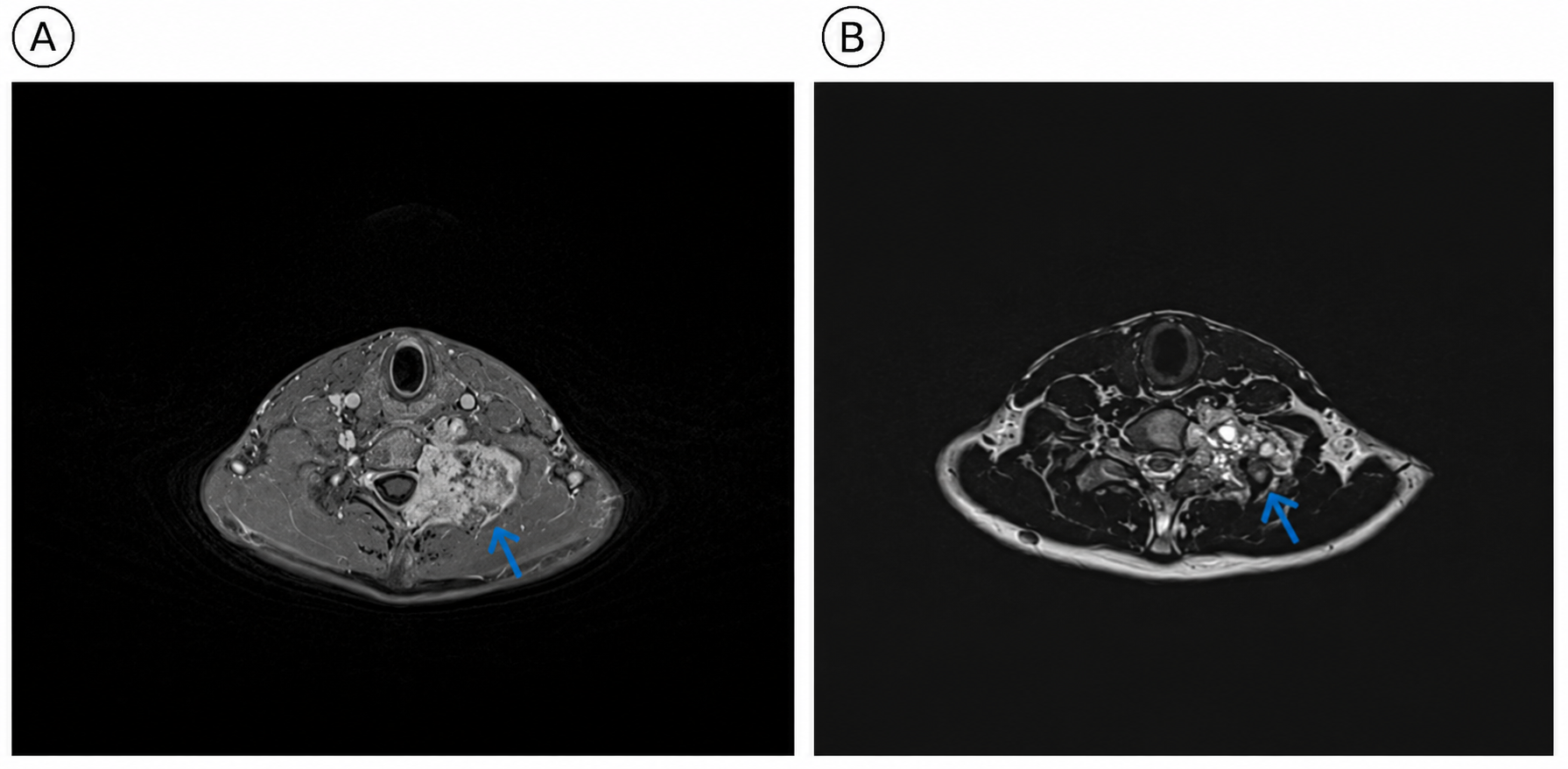
At one-month follow-up, the patient showed a marked response to physiotherapy: all provocative tests were relieved, scalene muscle tenderness had resolved, and the elevated arm stress test was negative, with only a mildly positive Elvey test on the affected side. A Doppler ultrasound performed for follow-up at this visit demonstrated a 40 × 15 × 10 mm heterogeneously hypoechoic left paraspinal lesion, suggesting compression of the adjacent nerve roots. For further characterization, magnetic resonance imaging (MRI) was obtained and revealed a well-circumscribed, expansile lytic mass at the C7 level measuring 55 × 36 × 32 mm (Figure 1).
Therapeutic Intervention
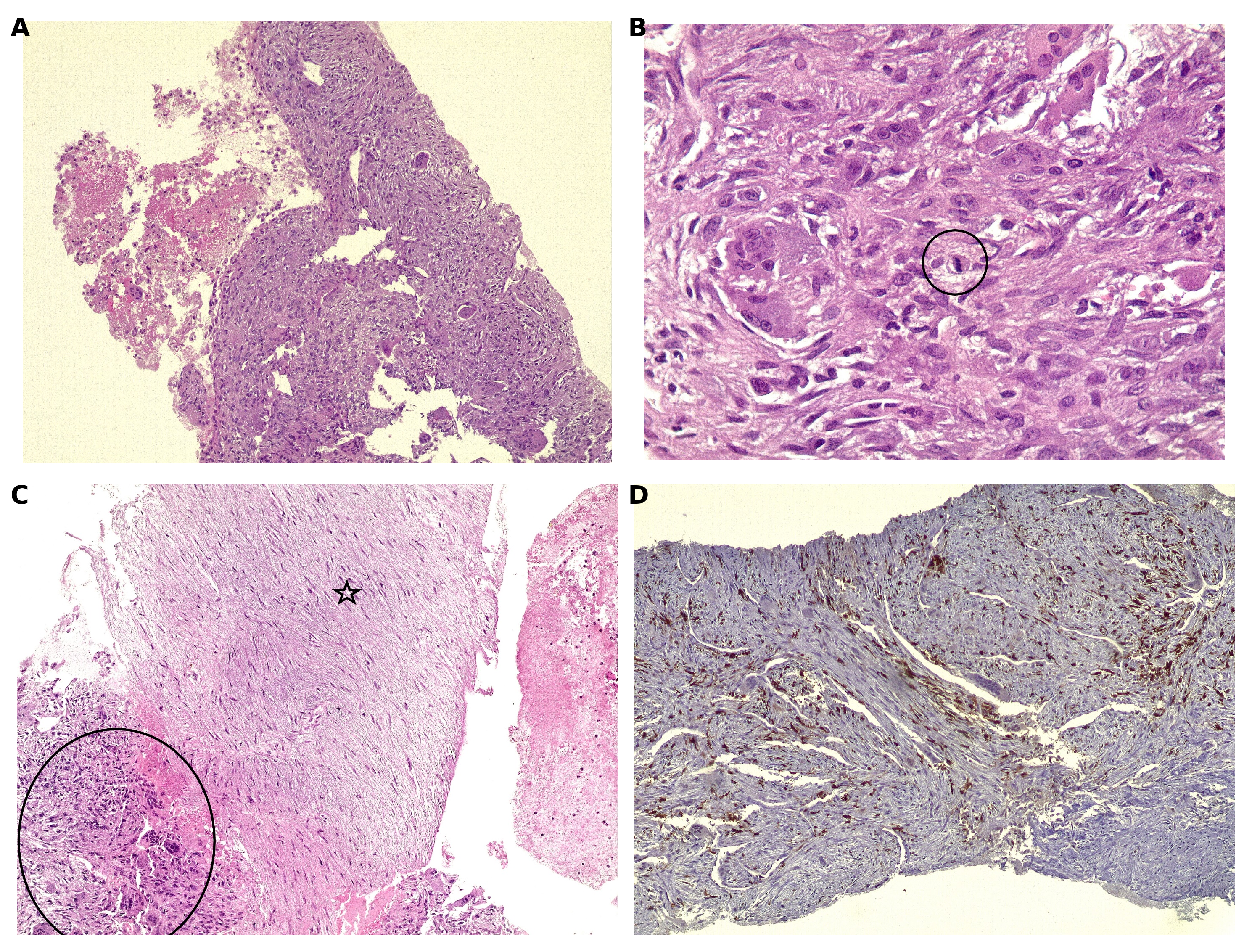
The patient was referred to the neurosurgery department for further work-up. A computed tomography (CT)-guided biopsy was scheduled, and biopsies were obtained from the left transverse process of the C7 vertebra using an 18-gauge coaxial needle. The procedure was uneventful, with no significant complications, and following it, the patient was able to move the left upper limb and hand without obvious limitation. Histopathological examination of the core biopsy showed blood-filled cystic spaces lined by fibrous septa, hypocellular myxoid foci with bland spindle cells, and fibroblastic areas containing unevenly distributed osteoclast-type giant cells, with no necrosis. Patchy strong cytoplasmic S100 staining supported chondroid differentiation. The lesion was reported as a giant cell-rich lesion with secondary aneurysmal bone cyst change, favoring chondromyxoid fibroma, with giant cell tumor of bone remaining in the differential (Figure 2).
Follow-up
Following multidisciplinary team discussion, surgical excision was recommended as definitive management because of the lesion’s location and potential for further enlargement, which could make subsequent excision more difficult, as well as the need for definitive histopathological diagnosis. However, the patient declined the proposed procedure and was subsequently lost to follow-up.
|
Peak (ms) |
Normal Peak (ms) |
P-T Amp (µV) |
Normal P-T Amp (µV) |
Site 1 |
Site 2 |
Delta-P (ms) |
Dist (cm) |
Vel (m/s) |
|
Left Medial Antebrachial Cutaneous Antidromic Sensory (Medial Forearm) |
||||||||
|
2.0 |
— |
13.0 |
— |
Elbow |
Medial Forearm |
2.0 |
0.0 |
— |
|
Right Medial Antebrachial Cutaneous Antidromic Sensory (Medial Forearm) |
||||||||
|
2.2 |
— |
18.1 |
— |
Elbow |
Medial Forearm |
2.2 |
0.0 |
— |
|
Left Median Antidromic Sensory (2nd Digit) |
||||||||
|
2.9 |
<3.5 |
150.6 |
>20 |
Wrist |
2nd Digit |
2.9 |
13.0 |
45 |
|
Left Ulnar Antidromic Sensory (5th Digit) |
||||||||
|
2.5 |
<3.1 |
94.2 |
>17.0 |
Wrist |
5th Digit |
2.5 |
11.0 |
44 |
|
Right Ulnar Antidromic Sensory (5th Digit) |
||||||||
|
2.9 |
<3.1 |
77.5 |
>17.0 |
Wrist |
5th Digit |
2.9 |
11.0 |
38 |
|
Abbreviations. P-T Amp, peak-to-peak amplitude; Delta-P, peak latency difference; Dist, distance; Vel, conduction velocity; ms, millisecond; µV, microvolt; cm, centimeter, m/s, meter per second |
||||||||
Discussion
Neurogenic TOS is the most difficult subtype to diagnose, as it lacks the objective vascular findings that help establish the venous and arterial forms. No single investigation is confirmatory, and the diagnosis therefore rests largely on clinical history and provocative maneuvers [3]. This challenge is compounded by the absence of a universally accepted diagnostic standard and by considerable symptom overlap with a wide range of musculoskeletal and neurological disorders, including cervical radiculopathy, carpal tunnel and ulnar entrapment syndromes, brachial plexus tumors, rotator cuff pathology, and Pancoast tumors [2]. Beyond these common mimics, clinicians should also remain alert to the rarer entities that can produce nTOS-like symptoms, namely discrete structural lesions of the cervicothoracic region, including osteoblastoma, anomalous muscles such as the subclavius posticus, space-occupying lesions such as lipomas, and post-surgical changes such as clavicular fixation screws, which are easily overlooked and frequently misdiagnosed [5]. The cases reviewed in the present report were all cervicothoracic lesions that presented as TOS despite an underlying structural pathology [10-14] (Table 2).
|
Author, year |
Age/Sex |
Presentation |
Lesion (histology) |
Site |
TOS subtype |
Diagnostic modality |
Mechanism |
Management & outcome |
Ref |
|
Present case, 2026 |
17/M |
Left shoulder pain, arm immobility (2 yr); positive Elvey & EAST |
Chondromyxo-id fibroma with aneurysmal bone cyst change |
C7 vertebr-a (body + left posteri-or elemen-ts) |
Neurogenic |
Doppler US, NCS, MRI, CT-guided biopsy |
Root-level mimic |
Surgery declined, lost to follow-up |
|
|
Medina, 2015 |
17/M |
Left-sided neck pain, hand numbness, weakness |
Aneurysmal bone cyst |
First rib (left) |
Neurogenic |
CT, MRI |
True outlet compression |
En bloc resection; resolved |
[10] |
|
Patel, 2016 |
55/M |
Right arm pain, paresthesia |
Chondrosarco-ma, grade I |
First rib (right) |
Neurogenic |
X-ray, CT, CT-guided biopsy |
True outlet compression |
Excision; no recurrence 21 months |
[11] |
|
Hamouri, 2021 |
23/F |
Cervical mass, pain, paresthesia |
Osteoblastom-a |
First rib (left) |
Neurogenic |
CT, MRI, NCS, incisional biopsy |
True outlet compression |
Excision; resolved, no recurrence 24 months |
[12] |
|
Singh, 2023 |
21/F |
Arm numbness, functional disability |
Aggressive epithelioid osteoblastoma |
Cervic-al rib (right) |
Neurogenic |
MRI, CT angiograph-y, CT-guided biopsy |
True outlet compression |
Excision + arterial graft; improved |
[13] |
|
Levy Faber, 2010 |
18/F |
Shoulder pain, arm pain, paresthesia |
Hydatid cyst (echinococcus-is) |
First rib (right, anterio-r) |
Combined |
X-ray, bone scan, CT, MRI; histopathol-ogy |
True outlet compression |
First-rib resection; resolved |
[14] |
|
Abbreviations. CT, computed tomography; EAST, elevated arm stress test; F, female; M, male; MRI, magnetic resonance imaging; NCS, nerve conduction study; TOS, thoracic outlet syndrome; US, ultrasound; Ref, Reference
|
|||||||||
The physical examination and provocative tests on which the diagnosis depends are of limited sensitivity and specificity. Maneuvers such as the elevated arm stress test, the upper limb tension (Elvey) test, and Adson's test are frequently positive in healthy individuals; used alone, their specificity is low (Adson's 76%, EAST 30%), rising to 82% only when combined [4]. Similarly, nerve conduction studies alone cannot establish a diagnosis of nTOS; a systematic review and meta-analysis found the evidence too heterogeneous and of too low quality to determine the sensitivity or specificity of electrophysiology, concluding that it serves mainly to exclude alternative conditions rather than to confirm the diagnosis [15]. Among the reviewed cases, pain was the most common symptom, with paresthesia, numbness, weakness, and functional impairment also reported [10-14]. In the present case, the patient was positive on both the Elvey and elevated arm stress tests, with reproducible symptoms and scalene tenderness. These findings, together with nerve conduction studies, were consistent with mild left-sided nTOS. Yet the same features could not distinguish outlet-level compression from a more proximal cause, and the true origin proved to be a structural lesion of the C7 vertebra rather than the outlet itself.
Therefore, imaging plays a central role in the evaluation of suspected nTOS, serving two complementary purposes: excluding the many conditions that mimic the syndrome and identifying any underlying structural lesion responsible for the symptoms. Because no single modality is both sensitive and specific for nTOS, these investigations are best used in combination rather than in isolation. Chest radiography, ideally with a dedicated thoracic aperture view, is a valuable initial study, allowing assessment of bony abnormalities such as cervical ribs and, though uncommonly, revealing an underlying tumor. It is complemented by MRI, which excludes cervical radiculopathy and characterizes soft-tissue and other structural abnormalities that may account for the presentation [6]. This diagnostic role of imaging is supported by the reviewed cases, in which the underlying lesion was identified only through cross-sectional and adjunctive imaging rather than clinical assessment alone. Across these reports, the responsible lesions were disclosed by combinations of plain radiography, CT, MRI, and, in several instances, image-guided biopsy [10-14]. In the present case, initial chest radiography was unremarkable, and it was only when a Doppler ultrasound performed at follow-up demonstrated a paraspinal lesion that the underlying cause became apparent; MRI then characterized a well-circumscribed expansile mass at C7, and CT-guided biopsy demonstrated a giant cell-rich lesion with secondary aneurysmal bone cyst change, favoring chondromyxoid fibroma. This progression illustrates that a normal initial radiograph does not exclude a structural cause of nTOS and that further cross-sectional imaging may be warranted when clinical findings remain unexplained.
All reviewed cases underwent surgical management tailored to the underlying lesion, including en bloc resection for aneurysmal bone cyst, excision for chondrosarcoma and osteoblastoma, excision with arterial grafting for cervical rib osteoblastoma, and first-rib resection for hydatid cyst. Complete symptom resolution was reported in 4/5 cases (80%), while clinical improvement was reported in 1/5 (20%). In the present case, surgery was recommended, but the patient declined the procedure and was subsequently lost to follow-up; therefore, treatment response and long-term outcome could not be assessed.
This report has several limitations. First, the patient declined the recommended surgical excision, so the definitive treatment of the lesion was never undertaken, and its management response could not be assessed. Second, he was subsequently lost to follow-up, leaving the natural history and long-term outcome of the condition unknown. Third, because surgery was not performed, the precise anatomical relationship between the C7 lesion and the affected nerve root was not confirmed operatively, and the proposed root-level mechanism, though consistent with the clinical and imaging findings, remains inferred rather than directly demonstrated.
Conclusion
Cervical vertebral lesions can mimic neurogenic thoracic outlet syndrome, highlighting the importance of imaging in identifying underlying structural pathology.
Declarations
Conflicts of Interest: The authors have no conflicts of interest to disclose.
Ethical Approval: Not applicable.
Consent for Participation: Not applicable.
Consent for Publication: Written informed consent was obtained from the patient's parent/legal guardian (the patient being a minor) for the publication of this case report and any accompanying images.
Funding: The present study received no financial support.
Acknowledgments: None to be declared.
Authors' Contributions: FHK conceived and designed the study, supervised the project, critically revised the manuscript, and was responsible for the overall accountability of the work. MIH contributed to the conception and design of the study and drafted the manuscript. SSM, RBJ, ASH, SKA, AKG, LAS, HHA, HAY, HSN, and HAA contributed to data acquisition, study design, and critical revision of the manuscript. All authors contributed to the interpretation of the findings, critically reviewed the manuscript, approved the final version, and agreed to be accountable for all aspects of the work. FHK and MIH confirm the authenticity of the data, and all authors approved the final manuscript.
Use of AI: Claude Opus 4.8 (Anthropic) was used to assist with grammar, writing, and paraphrasing. All final content was reviewed and approved by the authors.
Data Availability Statement: The data generated by this study are fully included in the figures and/or tables of this article.
References
- Peet RM, Henriksen JD, Anderson TP, Martin GM. Thoracic-Outlet Syndrome: Evaluation of a Therapeutic Exercise Program. Proc Staff Meet Mayo Clin. 1956;31(9):281-287.
- Kakamad FH, Asaad SK, Ghafour AK, Sabr NS, Namiq HS, Mustafa LJ, et al. Differential Diagnosis of Neurogenic Thoracic Outlet Syndrome: A Review. Barw Med J. 2025;3(2):36-44. doi:10.58742/bmj.v3i2.161
- Jones MR, Prabhakar A, Viswanath O, Urits I, Green JB, Kendrick JB, et al. Thoracic Outlet Syndrome: A Comprehensive Review of Pathophysiology, Diagnosis, and Treatment. Pain Ther. 2019;8(1):5-18. doi:10.1007/s40122-019-0124-2
- Povlsen S, Povlsen B. Diagnosing Thoracic Outlet Syndrome: Current Approaches and Future Directions. Diagnostics (Basel). 2018;8(1):21. doi:10.3390/diagnostics8010021
- Kakamad FH, Ghafour AK, Nasralla HA, Asaad SK, Sabr NS, Tahir SH, et al. Rare Etiologies of Thoracic Outlet Syndrome: A Systematic Review. World Acad Sci J. 2025;7(5):95. doi:10.3892/wasj.2025.383
- Teijink SBJ, Pesser N, Goeteyn J, Barnhoorn RJ, van Sambeek MRHM, van Nuenen BFL, et al. General Overview and Diagnostic (Imaging) Techniques for Neurogenic Thoracic Outlet Syndrome. Diagnostics (Basel). 2023;13(9):1625. doi:10.3390/diagnostics13091625
- Subach BR, Copay AG, Martin MM, Schuler TC, Romero-Gutierrez M. An Unusual Occurrence of Chondromyxoid Fibroma with Secondary Aneurysmal Bone Cyst in the Cervical Spine. Spine J. 2010;10(2):e5-e9. doi:10.1016/j.spinee.2009.11.016
- Prasad S, Nassar M, Azzam AY, García-Muro-San José F, Jamee M, Kasem Ali Sliman R, et al. CaReL Guidelines: A Consensus-Based Guideline on Case Reports and Literature Review (CaReL). Barw Med J. 2024;2(2):13-19. doi:10.58742/bmj.v2i2.89
- Kakamad FH, Mohammed SH, Abdalla BA, Hussein DA, Hammood ZD, Kakamad SH, et al. Non-Recommended Publishing Lists: Strategies for Detecting Deceitful Journals. Barw Med J. 2026;4(1):33-41. doi:10.58742/bmj.v4i1.227
- Medina M, Paul S. Aneurysmal Bone Cyst Arising from the First Rib: A Rare Cause of Thoracic Outlet Syndrome. Thorac Cardiovasc Surg Rep. 2015;5(1):74-76. doi:10.1055/s-0035-1569994
- Patel R, Devgarha S, Mathur RM. Chondrosarcoma of Right 1st Rib Presenting as Neurogenic Thoracic Outlet Syndrome; A 13th Case Report in World Literature. Egypt J Chest Dis Tuberc. 2016;65(3):707-709. doi:10.1016/j.ejcdt.2016.01.006
- Hamouri S, AlQudah M, Al-Zoubi N, Al Gargaz W, Jarboa' H, Hecker E. Rib Osteoblastoma as a Cause of Neurogenic Thoracic Outlet Syndrome: A Case Report. Am J Case Rep. 2021;22(1):e928548. doi:10.12659/AJCR.928548
- Singh D, Aryala S. An Unusual Case of Cervical Rib Tumor Presenting as Thoracic Outlet Syndrome. Indian J Vasc Endovasc Surg. 2023;10(1):68-71. doi:10.4103/ijves.ijves_27_22
- Levy Faber D, Best LA, Militianu D, Ben Nun A. Thoracic Outlet Syndrome Caused by Hydatid Cyst of the First Rib-Rare But Important. Indian J Surg. 2010;72(6):485-487. doi:10.1007/s12262-010-0126-0
- Daley P, Pomares G, Gross R, Menu P, Dauty M, Fouasson-Chailloux A. Use of Electroneuromyography in the Diagnosis of Neurogenic Thoracic Outlet Syndrome: A Systematic Review and Meta-Analysis. J Clin Med. 2022;11(17):5206. doi:10.3390/jcm11175206
Copyright (c) 2026 Fahmi H. Kakamad, Shakew S. Mohammed, Razan Babarasul Jalal, Azad S. Hattam, Saywan K. Asaad, Abdullah K. Ghafour, Lawand A. Sharif, Huzaifa Hamid Ahmed, Hadeel Adnan Yasseen, Miran I. Hassan, Hiwa S. Namiq, Harun Amanj Ahmed (Author)

This work is licensed under a Creative Commons Attribution 4.0 International License.